🧈
Pathology Lesson · Soft Tissue Tumour
Lipoma
Lesson 7 of 15 · Detailed pathology
PathologySoft Tissue Tumour
Points of Recognition
- 1Lobules of mature adipocytes
- 2Peripheral (compressed) nuclei
- 3Thin fibrous capsule
- 4Absence of atypia or lipoblasts
- 5Capillaries often present between adipocytes
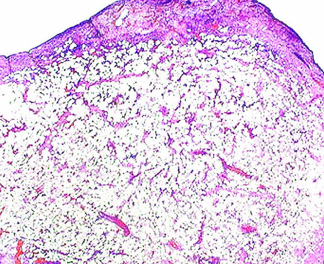
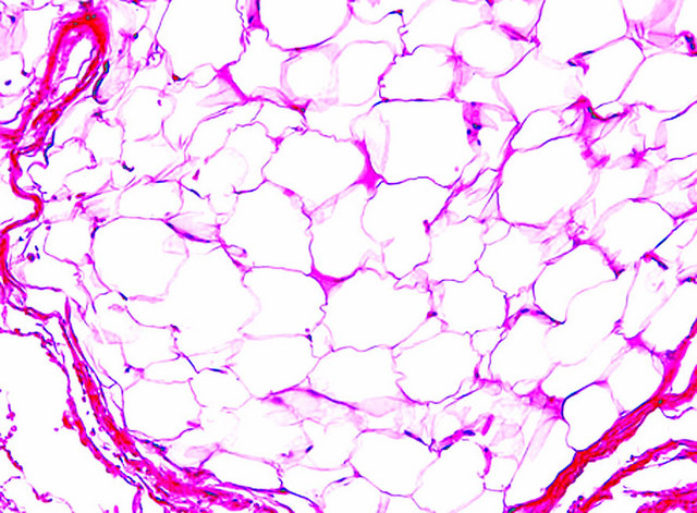
Image reference: PathologyOutlines.com
View topicDetailed Pathology
Definition
Lipoma is the most common benign soft-tissue tumour in adults, composed of mature adipocytes arranged in lobules separated by delicate fibrovascular septa. It is characterised by clear cytoplasm and peripherally displaced nuclei with no atypia, mitoses, or lipoblasts.
General / Essential Features
- Lobules of mature adipocytes with clear (lipid-filled) cytoplasm
- Peripherally located, small, flattened nuclei — no atypia or hyperchromasia
- Delicate fibrovascular septa separating lobules
- No lipoblasts (distinguish from well-differentiated liposarcoma)
- No increased mitotic activity
- May have thick fibrous capsule or blend imperceptibly into surrounding fat
Sites
- Subcutaneous tissue of trunk, shoulders, neck, and proximal extremities (most common)
- Intramuscular lipoma: deeper, less mobile, higher recurrence
- Spinal canal: epidural lipoma — compresses cord
- GI tract, breast, parotid (rare visceral sites)
Pathophysiology
Lipomas arise from clonal proliferation of mature adipocytes. Cytogenetic aberrations in chromosomes 12q, 13q, or 6p are common. The HMGA2 gene rearrangement on 12q15 is the most frequent molecular event. Angiolipomas contain a vascular component and are often multiple and painful.
Etiology
- Sporadic clonal mutations — most common
- Multiple hereditary lipomatosis: autosomal dominant
- Familial multiple lipomatosis: multiple lipomas often on forearms
- MEN1: parathyroid adenoma, pituitary adenoma, and lipomas
Clinical Features
- Soft, mobile, painless subcutaneous lump
- Slow growing; rarely exceeds 5 cm
- Angiolipoma: painful, especially after pressure
- Large intramuscular lipoma: firmness, pseudo-malignant feel
- No systemic features; malignant transformation is exceedingly rare
Diagnosis
- Clinical diagnosis: soft, mobile, painless lesion
- Ultrasound: hyperechoic or isoechoic lobulated lesion
- MRI: signal characteristics identical to normal fat — most reliable
- Excision biopsy and histopathology: definitive diagnosis
Treatment
- Observation if asymptomatic and <5 cm
- Surgical excision: for cosmetic reasons, pain, or diagnostic doubt
- Liposuction: alternative for superficial lipomas
- Intramuscular lipomas: wider excision to reduce recurrence
Video Lesson
References
- Kumar V, Abbas AK, Aster JC. Robbins & Cotran Pathologic Basis of Disease (10th ed.). Elsevier. 2020.
- Harsh Mohan. Textbook of Pathology (8th ed.). Jaypee Brothers. 2019.
- Bancroft JD, Layton C. Bancroft's Theory and Practice of Histological Techniques (8th ed.). Elsevier. 2019.
- PathologyOutlines.com. (2024). View topic
Ready to test yourself?
Apply what you've learned in the Pathology Spotting Test