logo
Pathology Lessons

Pathology

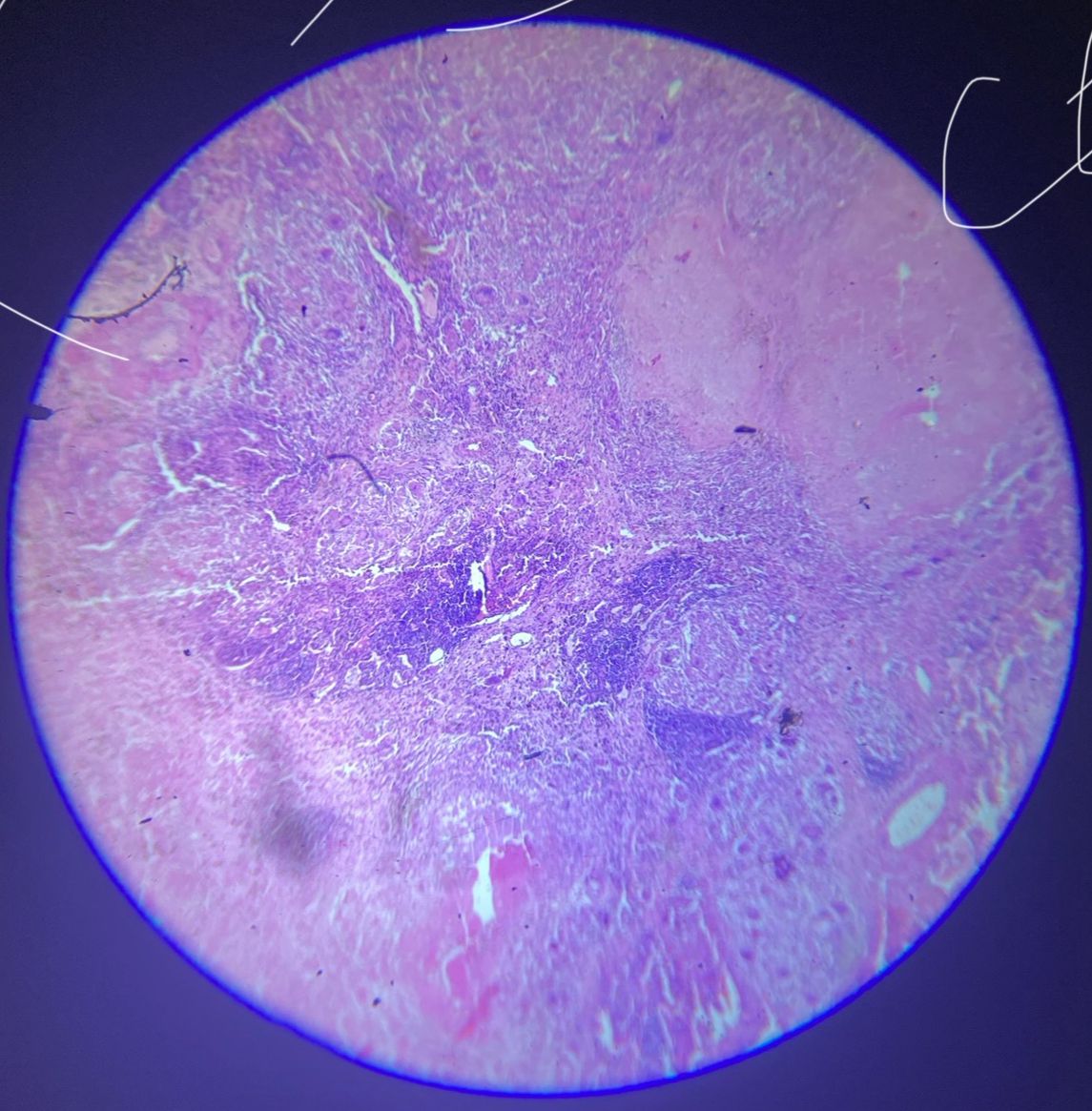
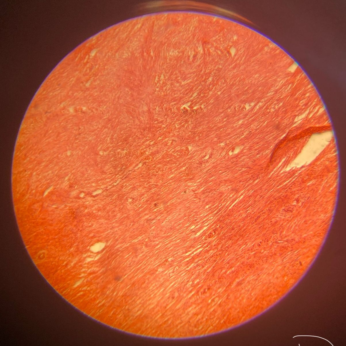
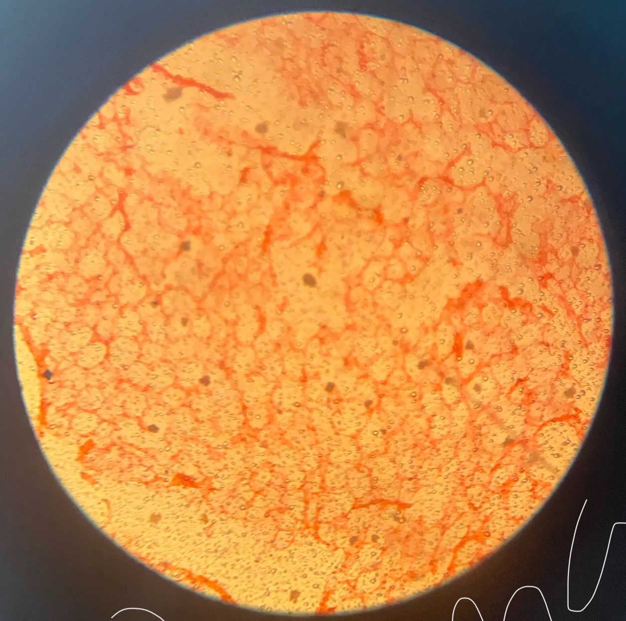
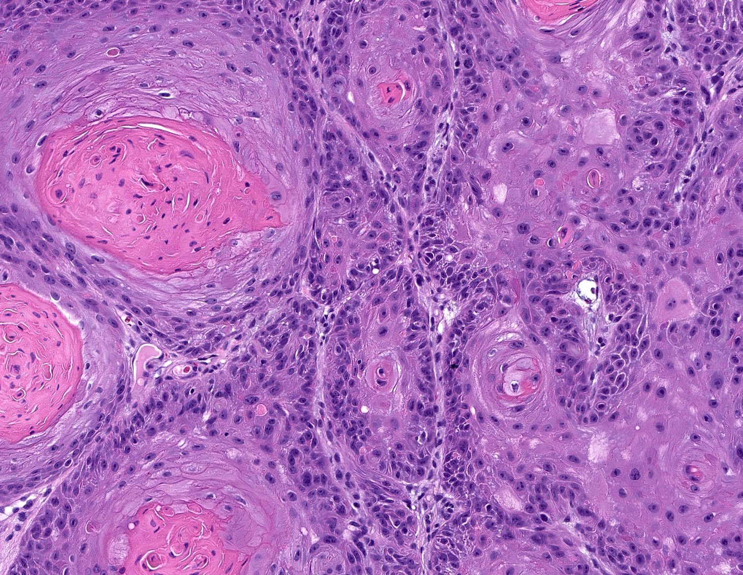
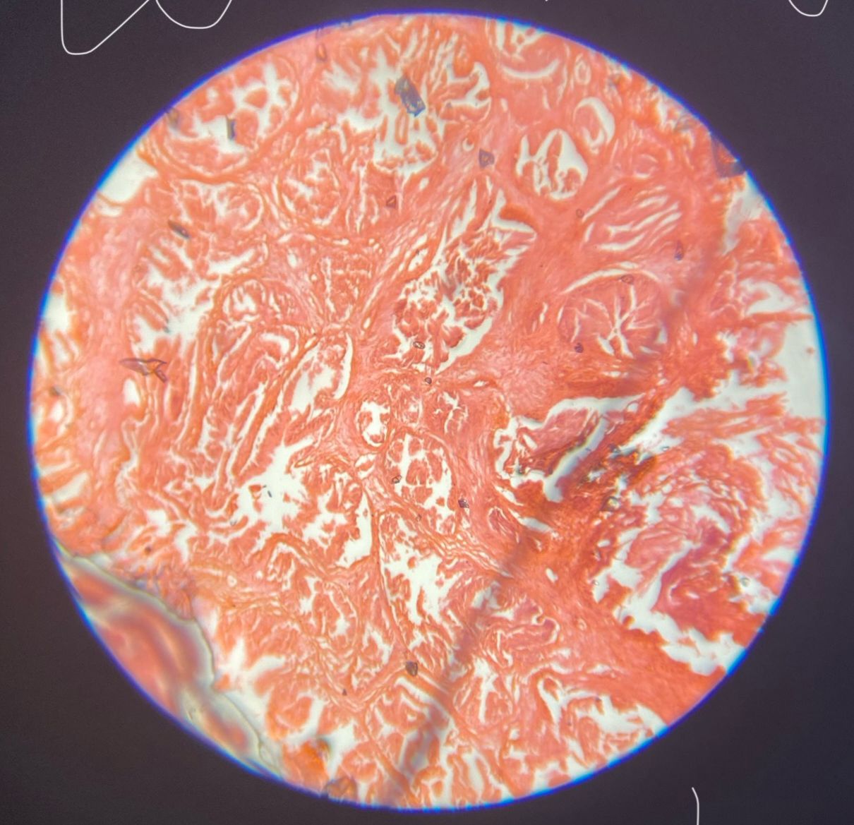
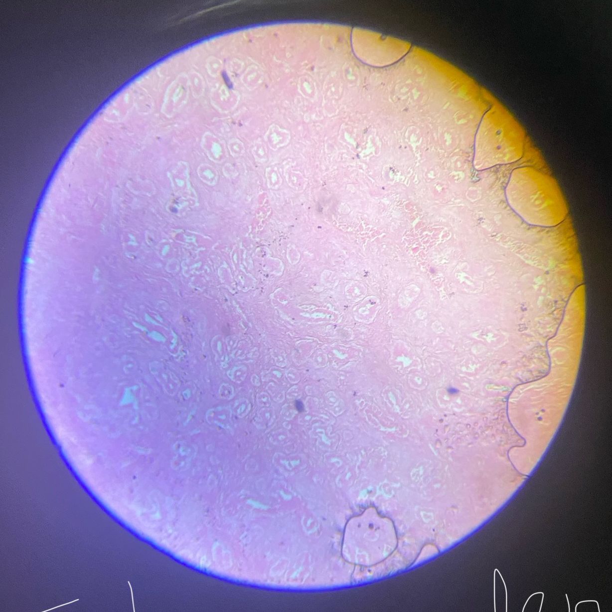
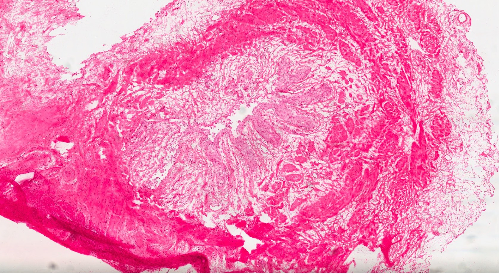
Disease Processes & Lesion Recognition

Examine each slide systematically — architecture first, then cellular detail — before reading the annotated lesson notes.

15
Lessons
4
Easy
7
Medium
4
Hard
Acute Appendicitis
Medium
1
GI Pathology

Acute Appendicitis

Transmural neutrophilic inflammation of the appendix — hyperplastic lymphoid follicles, luminal obstruction, peritoneal fibrin exudate.

8 min
Open
Chronic Cholecystitis
Medium
2
Hepatobiliary

Chronic Cholecystitis

Rokitansky–Aschoff sinuses, mononuclear infiltrate, sub-epithelial fibrosis, and smooth-muscle hypertrophy — almost always gallstone-associated.

8 min
Open
Gastritis
Easy
3
GI Pathology

Gastritis

Lymphocytic and neutrophilic mucosal inflammation with spiral H. pylori in the mucus layer — gastric pit distortion and intestinal metaplasia in chronic cases.

7 min
Open
Peptic Ulcer
Medium
4
GI Pathology

Peptic Ulcer

Full-thickness mucosal defect with four-zone base (exudate → necrosis → granulation → scar) — sharply punched-out edges, vessels at ulcer floor.

8 min
Open
TB Granuloma
Medium
5
Inflammatory

TB Granuloma

Central caseous necrosis, Langhans giant cells with horseshoe nuclei, epithelioid macrophages, lymphocytic rim — classic delayed hypersensitivity reaction.

9 min
Open
Leiomyoma
Medium
6
Smooth Muscle Tumour

Leiomyoma

Whorled fascicles of spindle smooth-muscle cells with cigar-shaped nuclei — commonest uterine neoplasm, oestrogen-dependent, low mitotic activity.

7 min
Open
Lipoma
Easy
7
Soft Tissue Tumour

Lipoma

Lobules of mature adipocytes with clear cytoplasm and peripheral nuclei, delicate fibrovascular septa — no atypia, no lipoblasts, no mitoses.

6 min
Open
Squamous Cell Carcinoma
Hard
8
Malignant Tumour

Squamous Cell Carcinoma

Keratin pearls, desmosomal bridges, polygonal eosinophilic cells with stromal invasion — arises from squamous epithelium or squamous metaplasia.

10 min
Open
Hodgkin's Disease
Hard
9
Haematological

Hodgkin's Disease

Pathognomonic Reed–Sternberg cells with owl-eye nucleoli (CD15+, CD30+) in a mixed inflammatory background — B-cell malignancy, bimodal age distribution.

11 min
Open
Adenocarcinoma
Hard
10
Malignant Tumour

Adenocarcinoma

Irregular malignant glands, abundant intraluminal mucin, invasion through muscularis mucosae, desmoplastic stroma — affects colon, stomach, lung, pancreas.

10 min
Open
Fatty Liver
Easy
11
Hepatic Pathology

Fatty Liver

Macro-vesicular steatosis with ballooned hepatocytes and dilated central vein — centrilobular distribution in NAFLD and alcoholic liver disease.

6 min
Open
Chronic Venous Congestion (Liver)
Medium
12
Hepatic Pathology

Chronic Venous Congestion (Liver)

Dilated central veins, centrilobular haemorrhagic necrosis, periportal fatty change — 'nutmeg liver' from right heart failure back-pressure.

7 min
Open
Benign Prostatic Hyperplasia
Medium
13
Urological

Benign Prostatic Hyperplasia

Cystic glands with papillary projections, double-layer epithelium (columnar + cuboidal basal), corpora amylacea — DHT-driven periurethral zone hyperplasia.

8 min
Open
Fibroadenoma
Easy
14
Breast Pathology

Fibroadenoma

Biphasic benign tumour with fibrous stroma compressing ducts into epithelial clefts — commonest breast tumour in women under 30, well-circumscribed.

6 min
Open
Carcinoma In Situ
Hard
15
Cervical Pathology

Carcinoma In Situ

Full-thickness cervical dysplasia with SMILE lesion — undifferentiated cells filling crypts, increased mitoses throughout, intact basement membrane.

9 min
Open
Ready to be tested?

Take the Pathology Spot Test — 15 shuffled slides, point-writing, instant scoring.

Start Spot Test