Stomach
Lesson 2 of 16 · Detailed theory + identification points
Points of Identification
4 pointsDetailed Theory
Object: Examination of Histological Slide of Stomach
General Overview
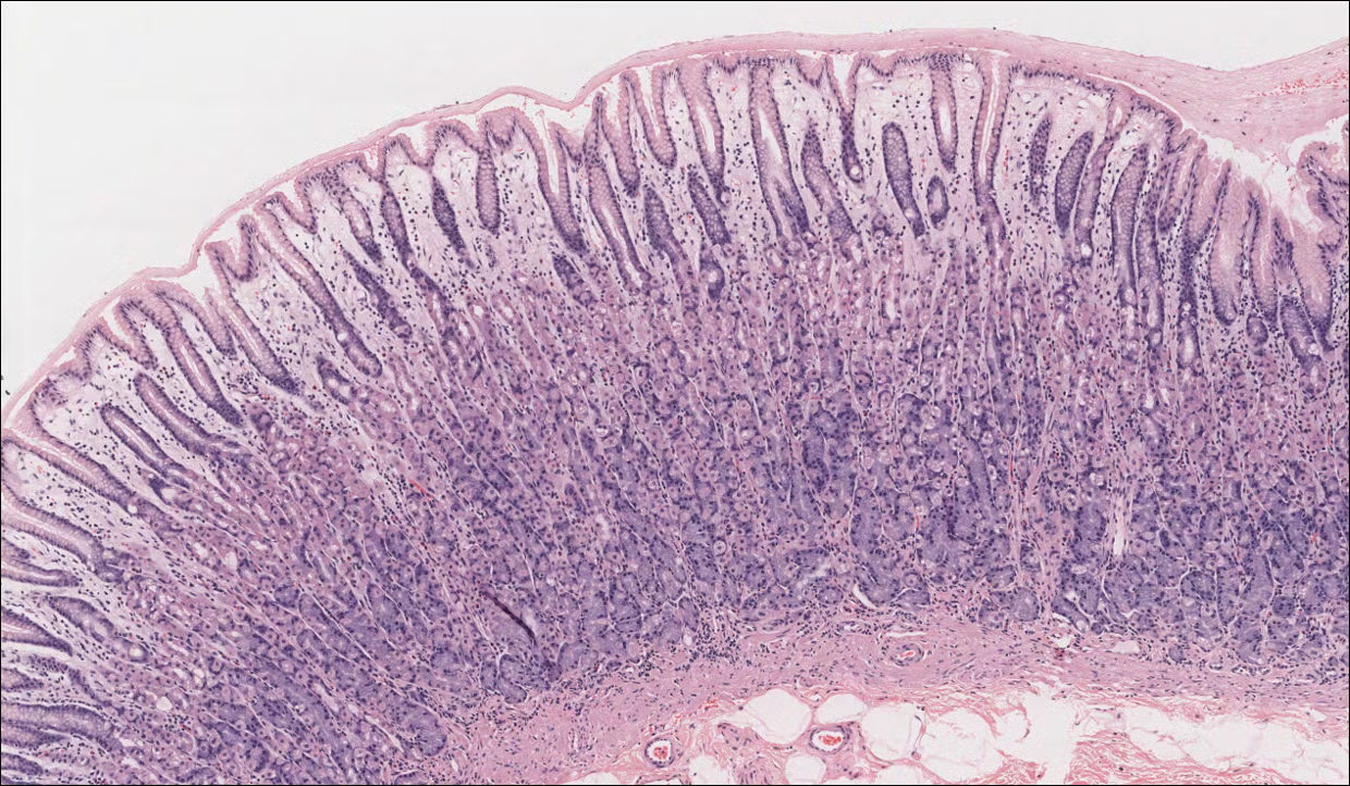
The stomach is a J-shaped muscular organ located between the oesophagus and the small intestine. It stores food, initiates protein digestion via HCl and pepsin, and regulates emptying into the duodenum. Its lumen is lined by a mucosa thrown into longitudinal folds called rugae that flatten when the stomach is distended.
Layers of the Stomach Wall
1. Mucosa
The innermost layer composed of: surface epithelium, lamina propria, and muscularis mucosae.
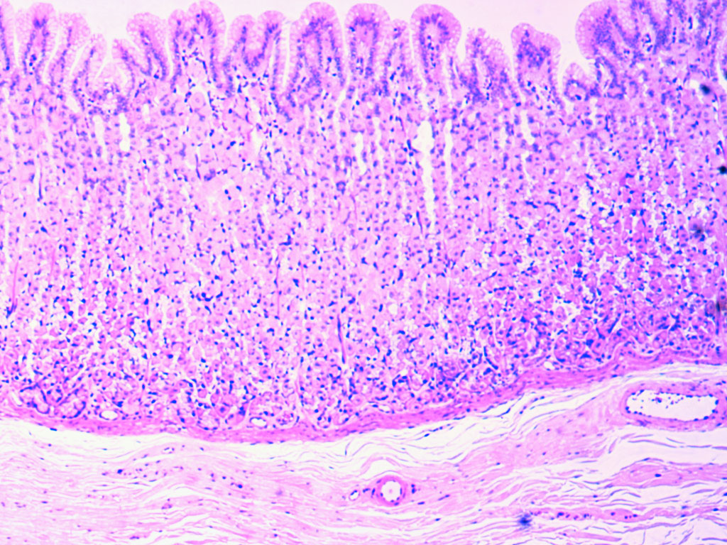
- Surface Epithelium: Simple columnar mucous epithelium. All cells are surface mucous cells secreting an alkaline mucus that protects the stomach from autodigestion.
- Gastric Pits (Foveolae Gastricae): Invaginations of the surface epithelium into the lamina propria. Each pit receives the openings of several gastric glands. Their depth varies by region — shallow in the fundus, deep in the antrum.
- Gastric Glands: Tubular glands in the lamina propria opening into the gastric pits. Three types by region:
2. Gastric Gland Cell Types (Fundic/Body Glands)
- Mucous Neck Cells: In the neck of the gland. Wedge-shaped with flattened basal nuclei and pale mucous apical cytoplasm. Secrete soluble mucus (different from surface mucus).
- Parietal (Oxyntic) Cells: Most numerous in the upper half of the gland. Large, pyramidal cells with deeply eosinophilic (pink) cytoplasm and centrally placed round nucleus. Contain intracellular canaliculi. Secrete HCl (via H⁺/K⁺-ATPase) and intrinsic factor (essential for vitamin B₁₂ absorption).
- Chief (Zymogenic) Cells: Predominate in the lower part of the gland. Basophilic cytoplasm (due to extensive rough ER), basally located nucleus. Secrete pepsinogen (activated to pepsin by HCl) and gastric lipase.
- Enteroendocrine Cells (APUD cells): Scattered throughout; secrete gastrin (G cells, antrum), somatostatin (D cells), histamine (ECL cells). Regulate acid secretion.
- Stem Cells: Located in the isthmus region; give rise to all other gland cell types.
3. Submucosa
Dense irregular connective tissue with blood vessels, lymphatics, and the submucosal (Meissner's) nerve plexus. No glands (unlike oesophagus and duodenum).
4. Muscularis Externa
Uniquely three-layered in the stomach: inner oblique, middle circular, outer longitudinal smooth muscle layers. This arrangement allows churning of food into chyme. The myenteric (Auerbach's) plexus lies between circular and longitudinal layers.
5. Serosa
Thin outer layer of loose connective tissue covered by visceral peritoneum (simple squamous mesothelium).
Regional Differences
- Cardia: Short cardiac glands, mainly mucous. Shallow gastric pits.
- Fundus and Body: Prominent gastric glands with all cell types. Pit:gland ratio ~1:4.
- Pyloric Antrum: Deep branched mucous glands, G cells present. Pyloric sphincter formed by thickened circular muscle.
Distinguishing Features on Slide
- Rugae (longitudinal folds) prominent at low magnification
- Gastric pits as regular invaginations of surface epithelium
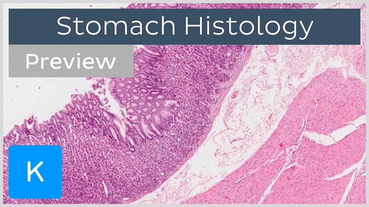
- Three-layered muscularis externa (key distinguishing feature from small intestine)
- No villi (distinguishes stomach from small intestine)
- Highly eosinophilic parietal cells prominent in fundic sections
References
5 sources- 1
Ross MH, Pawlina W. Histology: A Text and Atlas (8th ed.). Wolters Kluwer; 2020.
- 2
Young B, O'Dowd G, Woodford P. Wheater's Functional Histology (6th ed.). Churchill Livingstone/Elsevier; 2014.
- 3
Junqueira LC, Carneiro J. Basic Histology: Text & Atlas (13th ed.). McGraw-Hill; 2013.
- 4
Eroschenko VP. diFiore's Atlas of Histology with Functional Correlations (13th ed.). Wolters Kluwer; 2017.
- 5
Kumar V, Abbas AK, Aster JC. Robbins and Cotran Pathologic Basis of Disease (10th ed.). Elsevier; 2020.
Disclaimer: These notes are for educational purposes only and compiled from standard histology textbooks. Clinical interpretation of slides requires a qualified histologist or pathologist.
Ready to test yourself?
Apply what you've learned in the Histology Spotting Test