Kidney
Lesson 3 of 16 · Detailed theory + identification points
Points of Identification
4 pointsDetailed Theory
Object: Examination of Histological Slide of Kidney
General Overview
Kidneys are considered very important organs of the urinary system. They are enclosed in a thin connective tissue capsule. On their medial side there is a depression called the "Hilum" through which renal artery enters, and renal vein and ureter leave the organ.
Parts of Kidney
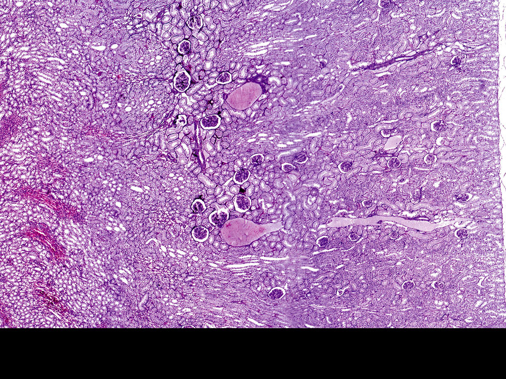
On cut section, the kidney consists of 2 parts:
- Cortex: The outer part of the kidney; granular in appearance. Includes the renal corpuscles, proximal convulated tubule (PCT), and distal convulated tubule (DCT). Cortical 80% consists of dilute urine, juxta-medullary, and concentrated urine.
- Medulla: The inner striated part of the kidney that includes the loop of Henle and the parallel blood vessels. The medulla is composed of 8–18 pyramids (conical masses) with base towards cortex and tip (papilla) towards pelvis. Each pyramid is a lobe of kidney along with its associated overlying cortex.
Renal Columns (of Bertin)
Extension of cortical substance into the medulla.
Medullary Rays
Extension of medullary substance into the cortex.
Stroma
Composed of scanty amount of interstitial connective tissue and blood vessels.
Parenchyma of the Kidney
Composed of Renal (Uriniferous) Tubules — they are of a large number and closely packed. Consist of 2 parts: (1) Nephron and (2) Collecting tubule.
1. Nephron
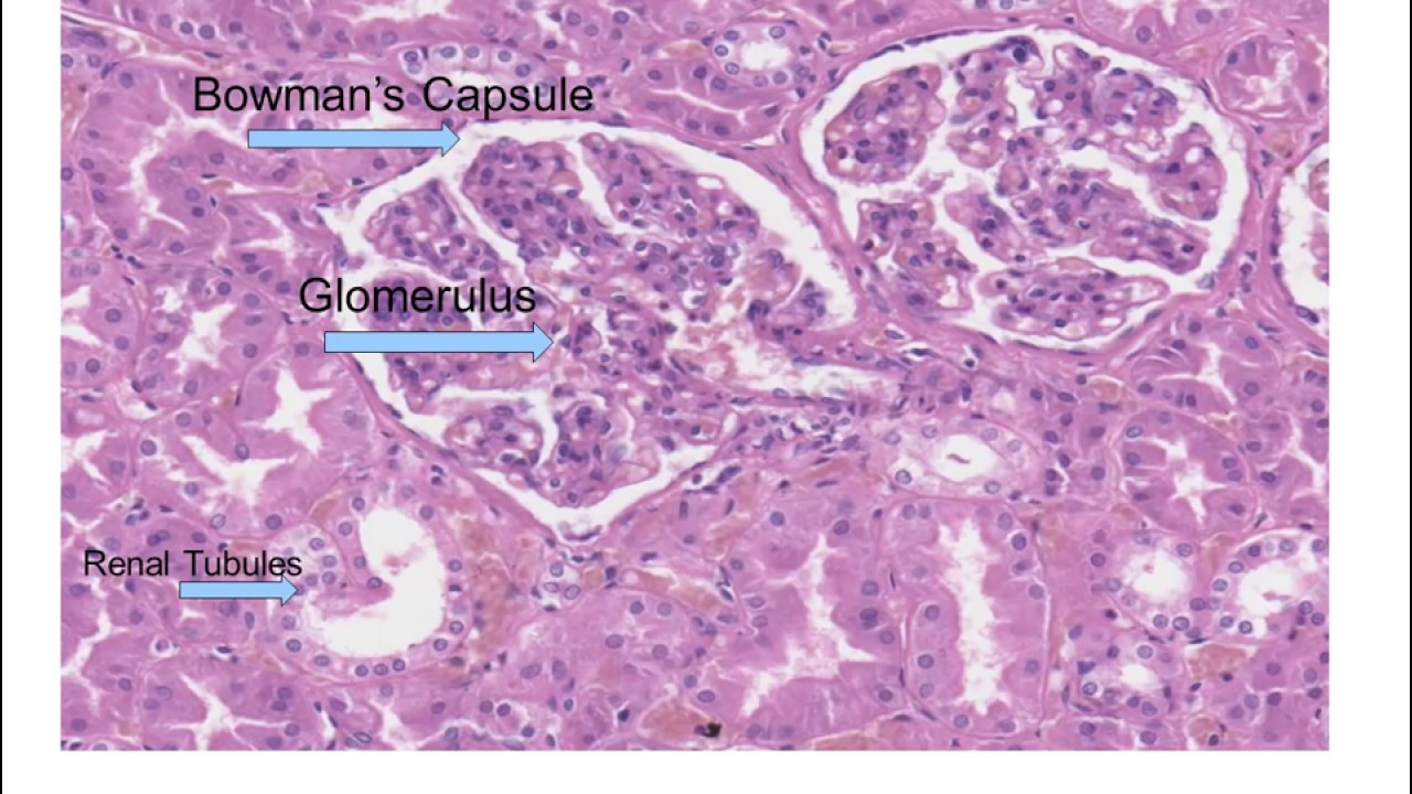
(a) Renal Corpuscle
Consists of glomerulus and Bowman's capsule.
Bowman's Capsule
Also called the glomerular capsule. It is a double-walled, cup-shaped dilation consisting of 2 layers:
- Outer (Parietal) Layer: Lined by simple squamous epithelium.
- Inner (Visceral) Layer: The visceral layer covers the glomerulus and invests it closely. It consists of a single layer of star-shaped epithelium cells called Podocytes. They share a common basal lamina with endothelial cells of glomerular capillaries.
Capsular / Urinary Space
The space that lies between the 2 layers mentioned above (parietal and visceral layer of Bowman's capsule). The cell bodies of podocytes are located in the capsular space. From the cell body arise primary processes that give rise to secondary processes or foot processes — "pedicels" — which make direct contact with the capsular surface of the common basal lamina.
Glomerulus
A tuft of capillaries lying inside the Bowman's capsule. It is composed of the following:
- (1) Afferent Arteriole: Entering the glomerulus.
- (2) Efferent Arteriole: Leaving the glomerulus.
- (3) Tuft of Capillaries.
- (4) Mesangium: Mesangial cells + extracellular matrix.
(b) Proximal Convulated Tubule (PCT)
The longest and most tortuous part of the renal tubule. Lined by columnar epithelial cells with a central nucleus and striated (brush) border — it has microvilli.
(c) Loop of Henle
Composed of the following segments:
- (1) Thick Descending Segment: Proximal straight tubule (PST) — similar to PCT.
- (2) Thin Ascending Segment: Lined by simple squamous cells.
- (3) Thin Descending Segment: Lined by simple squamous (flat) cells with central nucleus.
- (4) Thick Ascending Segment: Distal straight tubule (DST) — similar to DCT.
(d) Distal Convulated Tubule (DCT)
Shorter and less tortuous than PCT. Lined by simple cuboidal epithelium with no brush border.
Types of Nephron
There are 2 types based on the length of the loop of Henle:
- Cortical Nephron: Has a short loop of Henle.
- Juxta Glomerular or Medullary Nephron: Has a long loop of Henle extending at the cortico-medullary junction.
2. Collecting Tubules / Ducts
Excretory ducts — not a part of the nephron but connected to the distal convulated tubule by its short branches. They lie in the medullary rays. Lined by simple cuboidal cells with a central nucleus and no brush borders. They open into large papillary ducts of Bellini which open into the apex of renal papillae.
Filtration Barrier
Composed of the following:
- Fenestrated capillary endothelium.
- Basal lamina (common for both endothelial cells and podocytes).
- Filtration slits due to the presence of slit diaphragm.
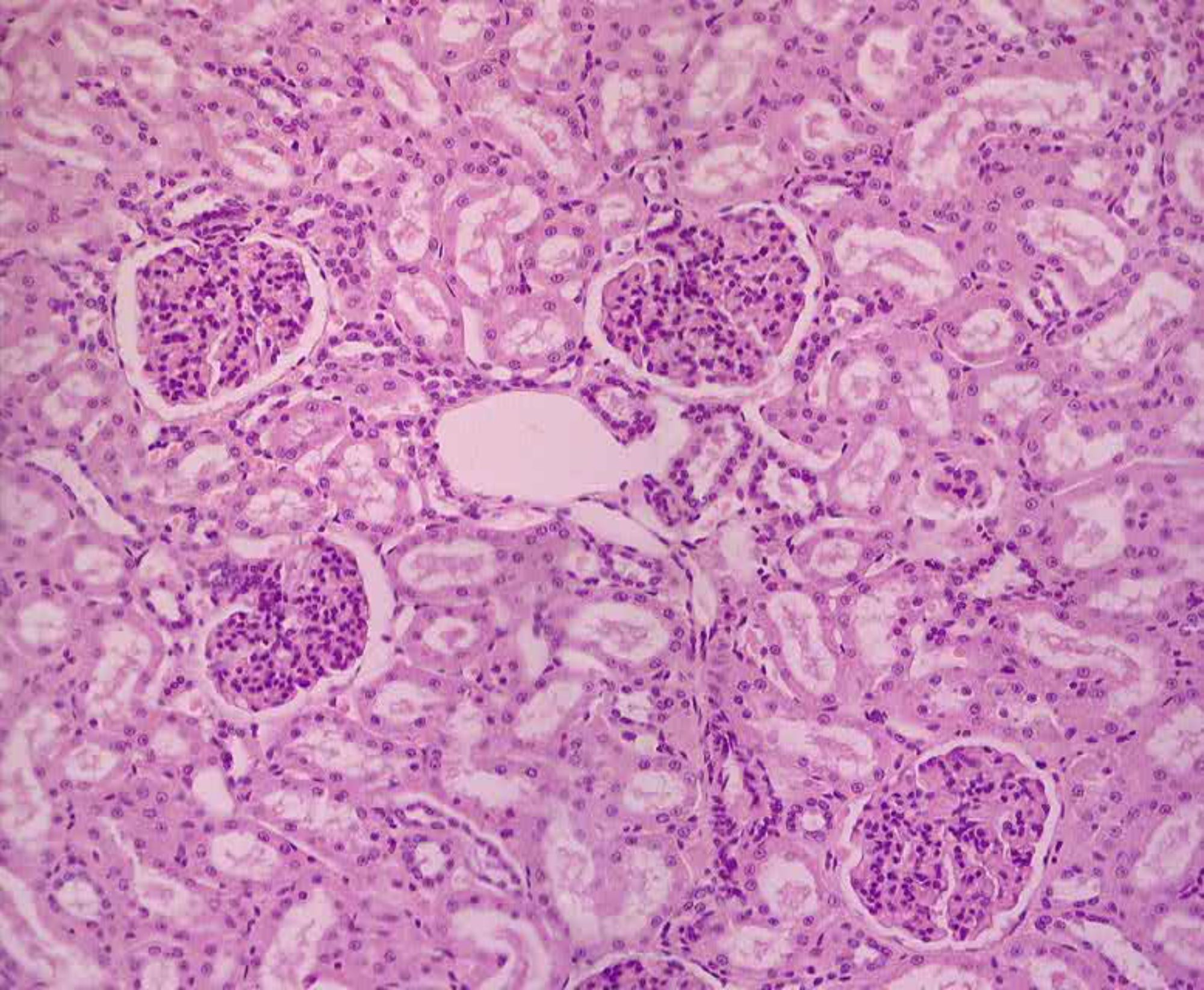
Distinguishing Features on Slide
- Renal corpuscles (glomerulus within Bowman's capsule) visible as round structures in cortex
- PCT with brush border and columnar cells — most prominent tubular profile
- DCT with cuboidal cells and no brush border — simpler epithelium
- Loop of Henle thin segments in medulla — lined by squamous cells
- Collecting ducts with cuboidal cells, very clear cell boundaries
- Rich blood vessel supply including afferent and efferent arterioles
References
5 sources- 1
Ross MH, Pawlina W. Histology: A Text and Atlas (8th ed.). Wolters Kluwer; 2020.
- 2
Young B, O'Dowd G, Woodford P. Wheater's Functional Histology (6th ed.). Churchill Livingstone/Elsevier; 2014.
- 3
Junqueira LC, Carneiro J. Basic Histology: Text & Atlas (13th ed.). McGraw-Hill; 2013.
- 4
Eroschenko VP. diFiore's Atlas of Histology with Functional Correlations (13th ed.). Wolters Kluwer; 2017.
- 5
Kumar V, Abbas AK, Aster JC. Robbins and Cotran Pathologic Basis of Disease (10th ed.). Elsevier; 2020.
Disclaimer: These notes are for educational purposes only and compiled from standard histology textbooks. Clinical interpretation of slides requires a qualified histologist or pathologist.
Ready to test yourself?
Apply what you've learned in the Histology Spotting Test