Simple Epithelium
Lesson 12 of 16 · Detailed theory + identification points
Points of Identification
5 pointsDetailed Theory
Object: Examination of Simple Epithelium
General Classification
Epithelium is classified by: (1) number of cell layers — simple (one layer) or stratified (multiple layers); and (2) shape of surface cells — squamous (flat), cuboidal, columnar, or transitional. Simple epithelium consists of a single layer of cells all directly attached to the basement membrane.
Mesothelium (serosa): flat cells with central bulging nuclei. Lines body cavities and vessels.
Thyroid follicles: cube-shaped cells with round, central nuclei. Secretes colloid.
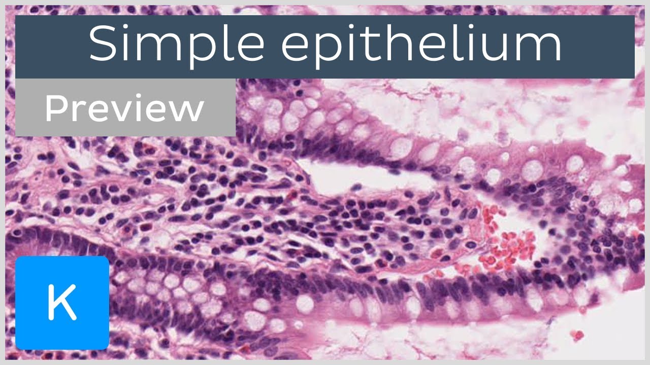
Intestine: tall cells with basal nuclei and apical brush border (microvilli). Goblet cells are also visible.
Trachea: nuclei at different levels (all cells touch basement membrane). Cilia and goblet cells present.
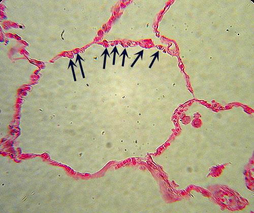
A — Simple Squamous Epithelium
Structure
Extremely thin, flat cells with a central disc-shaped or ovoid nucleus that bulges into the lumen. The cytoplasm is barely visible except around the nucleus. Cells fit together like floor tiles — irregular polygonal shapes with interlocking borders.
Locations and Function
- Mesothelium: lines all serous cavities (pleura, pericardium, peritoneum); reduces friction between organs
- Endothelium: lines all blood and lymphatic vessels; regulates exchange, haemostasis
- Alveoli (Type I pneumocytes): extremely thin for rapid gas diffusion
- Bowman's capsule parietal layer: kidney
- Loop of Henle (thin limbs): passive water/ion movement
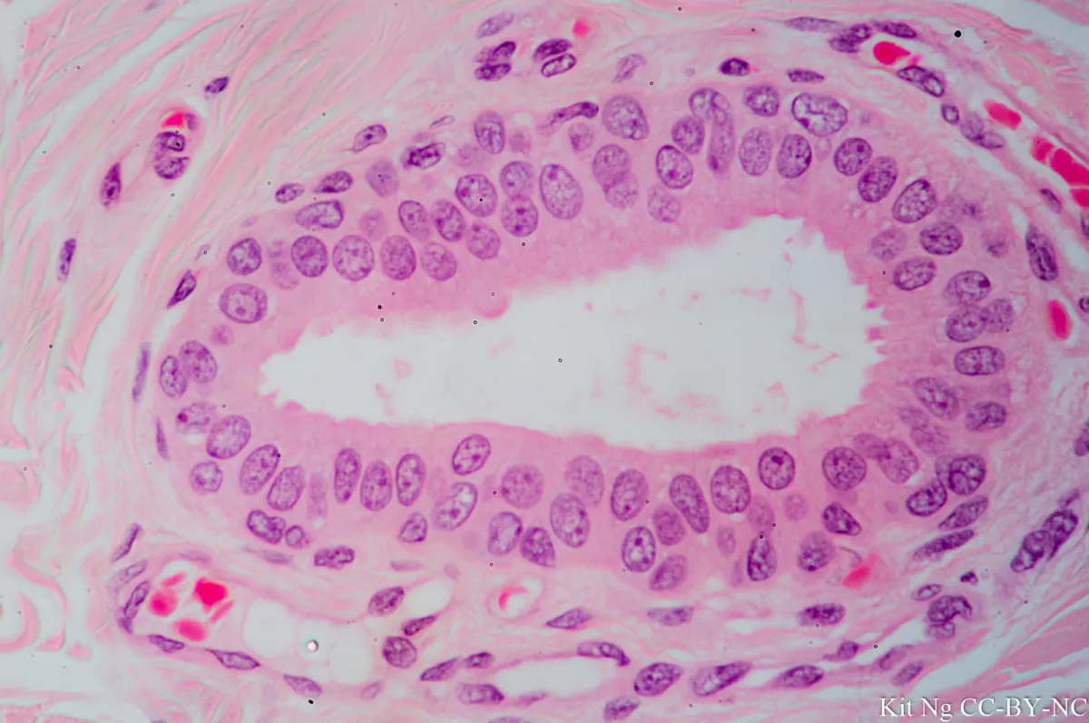
B — Simple Cuboidal Epithelium
Structure
Cells appear square in cross-section with a large, centrally placed spherical nucleus. Height approximately equals width. Mitochondria abundant in actively transporting cells.
Locations and Function
- Thyroid follicles: secrete thyroid hormone; height increases with TSH stimulation
- Kidney collecting tubules and distal convoluted tubule: reabsorption
- Small ducts of exocrine glands: pancreas, salivary glands
- Ovarian surface epithelium
- Choroid plexus of brain: produces CSF
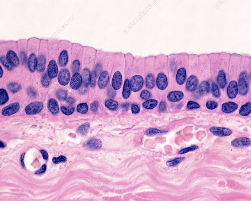
C — Simple Columnar Epithelium
Structure
Cells taller than they are wide; oval nuclei aligned in a row near the base. May possess: (a) microvilli (brush border) — in intestinal absorptive cells and PCT of kidney; (b) goblet cells — mucus-secreting unicellular glands interspersed among columnar cells; (c) cilia — in fallopian tube, uterus.
Locations and Function
- Stomach: surface mucous cells (no goblet cells, no brush border)
- Small intestine: enterocytes with brush border + goblet cells; absorption
- Large intestine: abundant goblet cells, fewer absorptive cells
- Gall bladder: simple tall columnar; no goblet cells; concentration of bile
- Fallopian tube: ciliated columnar — transport of ovum
- Endometrium
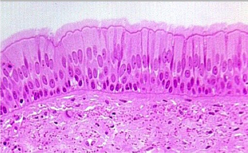
D — Pseudostratified Columnar Epithelium
Structure
Appears stratified because nuclei lie at different levels — but all cells contact the basement membrane (confirmed by EM). It is technically simple epithelium. Most commonly ciliated with goblet cells (respiratory epithelium).
- Trachea, bronchi, nasal cavity, paranasal sinuses: pseudostratified ciliated columnar with goblet cells (respiratory epithelium). Cilia beat synchronously to move the mucus blanket (mucociliary escalator) upward.
- Epididymis: pseudostratified with stereocilia (non-motile, long microvilli) — for reabsorption
- Male urethra (part)
Key Distinguishing Feature
All nuclei seem to be at different levels but no free surface cells are uninucleated — every nucleus is connected to the basement membrane if traced.
References
4 sources- 1
Ross MH, Pawlina W. Histology: A Text and Atlas (8th ed.). Wolters Kluwer; 2020.
- 2
Young B, O'Dowd G, Woodford P. Wheater's Functional Histology (6th ed.). Churchill Livingstone/Elsevier; 2014.
- 3
Junqueira LC, Carneiro J. Basic Histology: Text & Atlas (13th ed.). McGraw-Hill; 2013.
- 4
Eroschenko VP. diFiore's Atlas of Histology with Functional Correlations (13th ed.). Wolters Kluwer; 2017.
Disclaimer: These notes are for educational purposes only and compiled from standard histology textbooks. Clinical interpretation of slides requires a qualified histologist or pathologist.
Ready to test yourself?
Apply what you've learned in the Histology Spotting Test