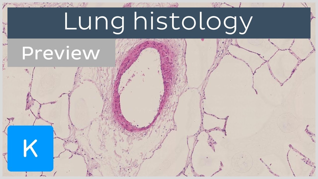
Lungs
Lesson 1 of 16 · Detailed theory + identification points
Points of Identification
4 pointsDetailed Theory
Object: Examination of Histological Slide of Lung
General Overview
The lungs are the primary organs of the respiratory system, responsible for gas exchange between atmospheric air and the bloodstream. Each lung is divided into lobes — right into three, left into two — separated by fissures and enclosed within a pleural sac. The hilum is where bronchi, pulmonary vessels, lymphatics, and nerves enter and exit.
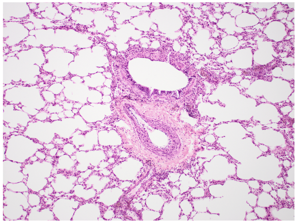
Histological Organisation
1. Conducting Zone
Begins at the trachea and extends through bronchi, bronchioles, and terminal bronchioles. No gas exchange occurs here; its function is to warm, humidify, and filter inspired air.
- Trachea and Primary Bronchi: Lined by pseudostratified ciliated columnar epithelium (respiratory epithelium) with goblet cells. Supported by C-shaped hyaline cartilage rings.
- Secondary and Tertiary Bronchi: Cartilage becomes irregular plates; smooth muscle increases. Submucosal mixed seromucous glands present.
- Bronchioles (<1 mm diameter): No cartilage. Simple ciliated columnar to cuboidal epithelium. Club (Clara) cells appear — dome-shaped non-ciliated secretory cells producing surfactant components and detoxifying enzymes.
- Terminal Bronchioles: Last purely conducting segment. Ciliated cells diminish; club cells predominate.
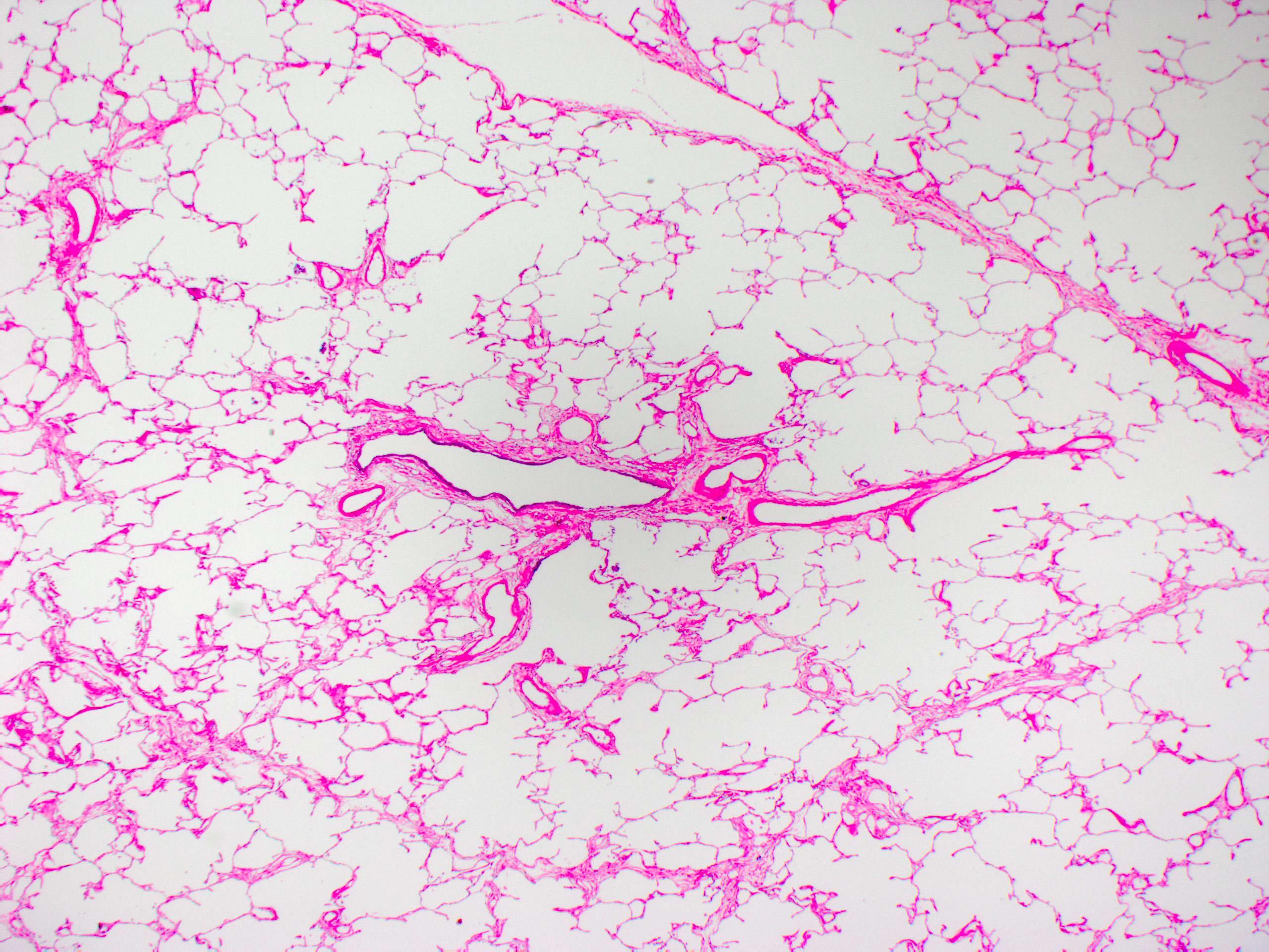
2. Respiratory Zone
- Respiratory Bronchioles: Walls interrupted by outpouching alveoli. Lined by simple cuboidal epithelium with occasional cilia.
- Alveolar Ducts: Long passages with walls entirely composed of alveolar openings. Smooth muscle at alveolar rims.
- Alveolar Sacs: Two or more alveoli sharing a common opening.
- Alveoli: Primary sites of gas exchange. Thin-walled, polyhedral sacs ~200 µm diameter, separated by interalveolar septa.
3. Alveolar Cells (Pneumocytes)
- Type I Pneumocytes: Cover ~95% of alveolar surface. Extremely thin (0.2 µm) flat cells allowing rapid gas diffusion. Cannot divide.
- Type II Pneumocytes: Cuboidal cells at alveolar corners with lamellar bodies. Secrete surfactant (dipalmitoyl phosphatidylcholine) — reduces surface tension, prevents alveolar collapse. Can divide and differentiate into Type I after injury.
- Alveolar Macrophages (Dust Cells): Phagocytic monocyte-derived cells in alveolar lumen and interstitium. Remove inhaled particles and microbes.
4. Blood–Air Barrier
O₂ and CO₂ diffuse across: cytoplasm of Type I pneumocyte → fused basal laminae → cytoplasm of capillary endothelial cell. Total thickness ~0.5 µm for rapid diffusion.
5. Pulmonary Vasculature
Pulmonary arteries (deoxygenated blood) travel alongside bronchi and bronchioles. Pulmonary veins travel in interlobular septa. Bronchial arteries supply bronchial walls with oxygenated blood. Rich lymphatics drain towards the hilum.
6. Pleura
Both visceral and parietal pleurae lined by a single layer of mesothelial cells (simple squamous epithelium) overlying connective tissue rich in elastic fibres, collagen, and blood/lymph vessels.
Distinguishing Features on Slide
- Abundant thin-walled air spaces giving a sponge-like appearance
- Bronchi/bronchioles as round/oval structures with prominent smooth muscle walls
- Distinct pulmonary blood vessels (arteries thick-walled, veins thinner)
- Interalveolar septa containing capillaries
- Scattered alveolar macrophages in alveolar spaces
References
5 sources- 1
Ross MH, Pawlina W. Histology: A Text and Atlas (8th ed.). Wolters Kluwer; 2020.
- 2
Young B, O'Dowd G, Woodford P. Wheater's Functional Histology (6th ed.). Churchill Livingstone/Elsevier; 2014.
- 3
Junqueira LC, Carneiro J. Basic Histology: Text & Atlas (13th ed.). McGraw-Hill; 2013.
- 4
Eroschenko VP. diFiore's Atlas of Histology with Functional Correlations (13th ed.). Wolters Kluwer; 2017.
- 5
Kumar V, Abbas AK, Aster JC. Robbins and Cotran Pathologic Basis of Disease (10th ed.). Elsevier; 2020.
Disclaimer: These notes are for educational purposes only and compiled from standard histology textbooks. Clinical interpretation of slides requires a qualified histologist or pathologist.
Ready to test yourself?
Apply what you've learned in the Histology Spotting Test