Stratified Epithelium
Lesson 13 of 16 · Detailed theory + identification points
Points of Identification
5 pointsDetailed Theory
Object: Examination of Stratified Epithelium
General Principle
Stratified epithelium has multiple cell layers — only the basal layer contacts the basement membrane. The surface cell shape defines the type. Primary function: protection from mechanical abrasion, chemical injury, and desiccation.
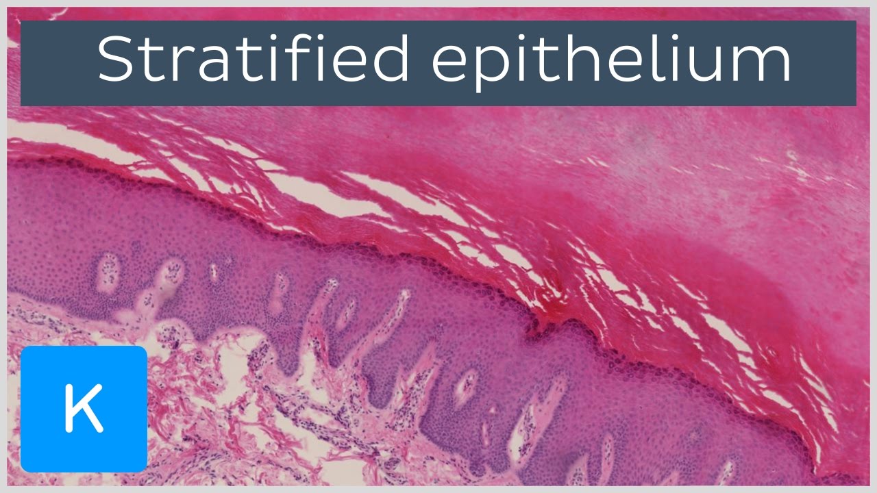
Oesophagus: multiple layers, surface cells flattened but nucleated.
Sweat gland duct: 2–3 layers of cuboidal cells lining the lumen.
Large excretory duct: surface columnar cells overlying cuboidal basal layers.
Urinary bladder (relaxed): dome-shaped umbrella cells, intermediate layers.
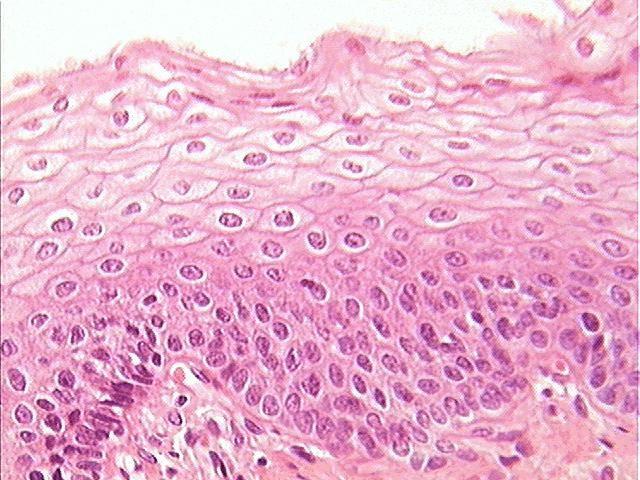
A — Stratified Squamous Epithelium
Non-Keratinised
Surface cells are flattened but retain their nuclei and remain viable. Found where a moist, smooth surface is needed: oral cavity, pharynx, oesophagus, vagina, ectocervix, conjunctiva. Layers:
- Stratum basale: columnar/cuboidal, mitotically active
- Stratum spinosum: polygonal cells with desmosomes
- Surface: flat nucleated squames
Keratinised (Epidermis)
Found only in skin. Surface cells are anucleate, filled with hard keratin. Five layers: stratum basale, spinosum, granulosum, lucidum (thick skin only), corneum. Provides a complete waterproof barrier.
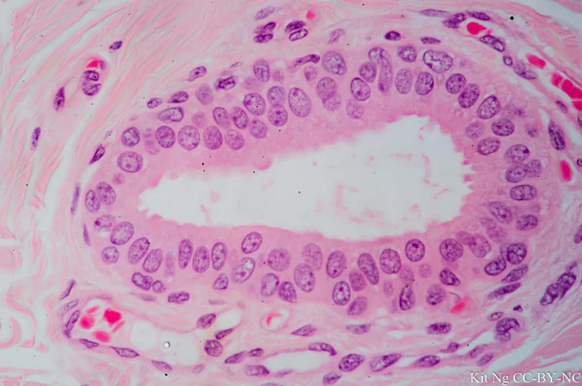
B — Stratified Cuboidal Epithelium
Two or more layers of cuboidal cells. Rare — found in large ducts of exocrine glands (sweat gland ducts, salivary gland ducts). Provides mechanical protection for duct walls while allowing secretion transport.
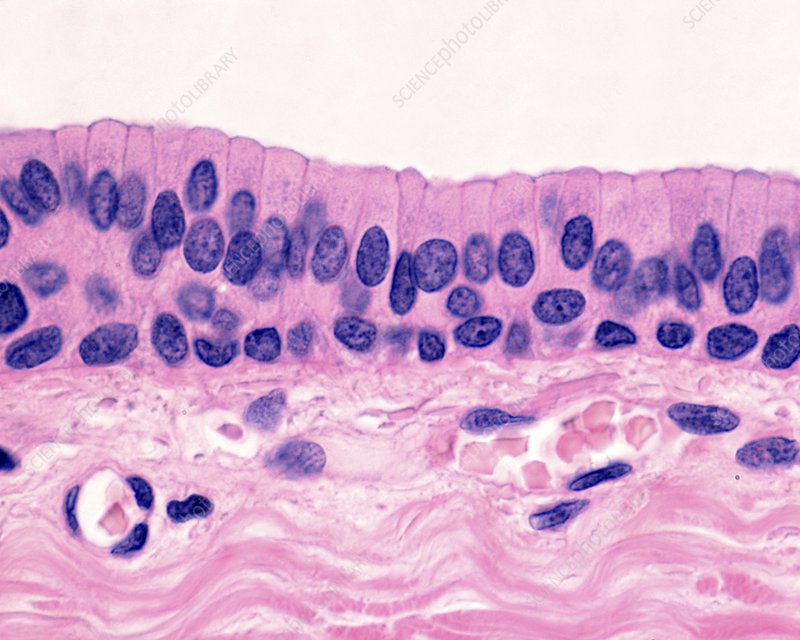
C — Stratified Columnar Epithelium
Multiple layers; surface cells are columnar. Very rare — found in largest ducts of mammary glands, male urethra (membranous), and conjunctiva near lid margin. Often transitional between other epithelial types.
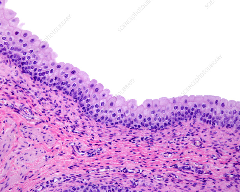
D — Transitional Epithelium (Urothelium)
Structure
Unique to the urinary tract (renal pelvis, ureter, urinary bladder, proximal urethra). Designed to accommodate stretch and recoil. Three layers:
- Basal layer: small cuboidal cells on basement membrane
- Intermediate layer: polygonal cells, 1–3 layers depending on distension
- Superficial layer (umbrella/facet cells): large, dome-shaped cells with a thick apical plasma membrane (asymmetric unit membrane — AUM) resistant to hypertonic urine; may be binucleate
Relaxed vs Distended States
- Relaxed (empty bladder): appears thick — 6–8 cell layers; cells rounded; umbrella cells dome-shaped
- Distended (full bladder): cells flatten; appears only 2–3 layers; umbrella cells spread flat
Key Histological Features
- Dome-shaped umbrella cells — most distinctive feature
- Thick apical membrane (AUM) visible at EM
- Underlying lamina propria rich in elastic fibres (allows recoil)
- No goblet cells, no brush border
References
4 sources- 1
Ross MH, Pawlina W. Histology: A Text and Atlas (8th ed.). Wolters Kluwer; 2020.
- 2
Young B, O'Dowd G, Woodford P. Wheater's Functional Histology (6th ed.). Churchill Livingstone/Elsevier; 2014.
- 3
Junqueira LC, Carneiro J. Basic Histology: Text & Atlas (13th ed.). McGraw-Hill; 2013.
- 4
Eroschenko VP. diFiore's Atlas of Histology with Functional Correlations (13th ed.). Wolters Kluwer; 2017.
Disclaimer: These notes are for educational purposes only and compiled from standard histology textbooks. Clinical interpretation of slides requires a qualified histologist or pathologist.
Ready to test yourself?
Apply what you've learned in the Histology Spotting Test