Skeletal Muscle
Lesson 8 of 16 · Detailed theory + identification points
Points of Identification
4 pointsDetailed Theory
Object: Examination of Histological Slide of Skeletal Muscle
General Overview
Skeletal muscle is voluntary, striated muscle attached to the skeleton via tendons. It accounts for ~40% of body weight. Each skeletal muscle fibre is a syncytium formed by the fusion of many myoblasts during development — hence it is multinucleated.
Organisation (from largest to smallest)
- Whole Muscle: Enclosed by epimysium (dense connective tissue).
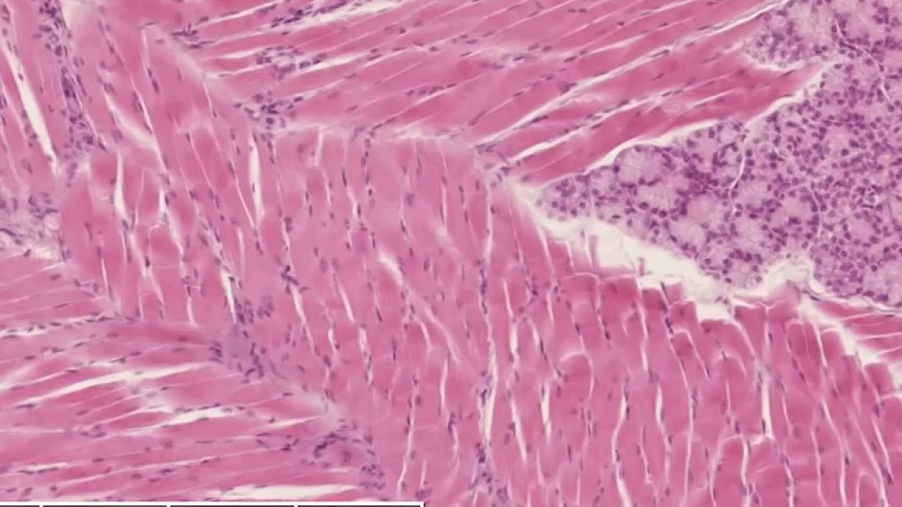
- Fascicle: Bundle of muscle fibres enclosed by perimysium.
- Muscle Fibre (Cell): Individual multinucleated cell enclosed by endomysium (fine reticular fibres + basal lamina).
- Myofibril: Contractile unit within each fibre; runs parallel to the long axis.
- Sarcomere: Repeating functional unit of the myofibril (Z-line to Z-line).
The Sarcomere — Basis of Striations
1. A Band (Anisotropic — Dark)
Contains thick myosin filaments (+ overlapping actin). Appears dark (anisotropic in polarised light). The A band does NOT shorten during contraction.
2. I Band (Isotropic — Light)
Contains only thin actin filaments. Appears light. The I band SHORTENS during contraction as actin slides over myosin.
3. H Zone
The central pale region of the A band — contains only myosin (no actin overlap). Narrows/disappears during contraction.
4. M Line
Runs through the centre of the H zone. Cross-links myosin filaments.
5. Z Line (Z Disc)
Bisects the I band. Actin filaments of adjacent sarcomeres are anchored here. The distance between two Z lines = one sarcomere (~2.5 µm at rest).
Cell Features
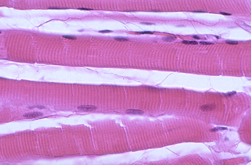
Peripheral Nuclei
Multiple nuclei are pushed to the periphery (sub-sarcolemmal), just beneath the cell membrane. This contrasts with cardiac muscle (central nuclei) and smooth muscle (central nuclei). A fibre may have hundreds of nuclei per centimetre of length.
Sarcoplasm and Organelles
The sarcoplasm contains abundant mitochondria (between myofibrils and beneath the sarcolemma), glycogen granules, and lipid droplets. The extensive sarcoplasmic reticulum forms a network around each myofibril for Ca²⁺ storage and release.
T-Tubules
Invaginations of the sarcolemma at the A-I junctions (two per sarcomere). Together with lateral cisternae of the SR, they form a triad — the functional unit for excitation-contraction coupling.
Fibre Types
- Type I (Slow-twitch, Red): Oxidative metabolism; many mitochondria and myoglobin; fatigue-resistant. Postural muscles.
- Type IIa (Fast-twitch, Red): Oxidative + glycolytic; intermediate fatigability.
- Type IIx/IIb (Fast-twitch, White): Glycolytic; few mitochondria; fast but fatigable. Sprinting muscles.
Neuromuscular Junction (Motor End Plate)
The terminal branching of a motor neuron forms synapses on specialised regions of the sarcolemma with junctional folds that increase surface area for ACh receptors (nAChR).
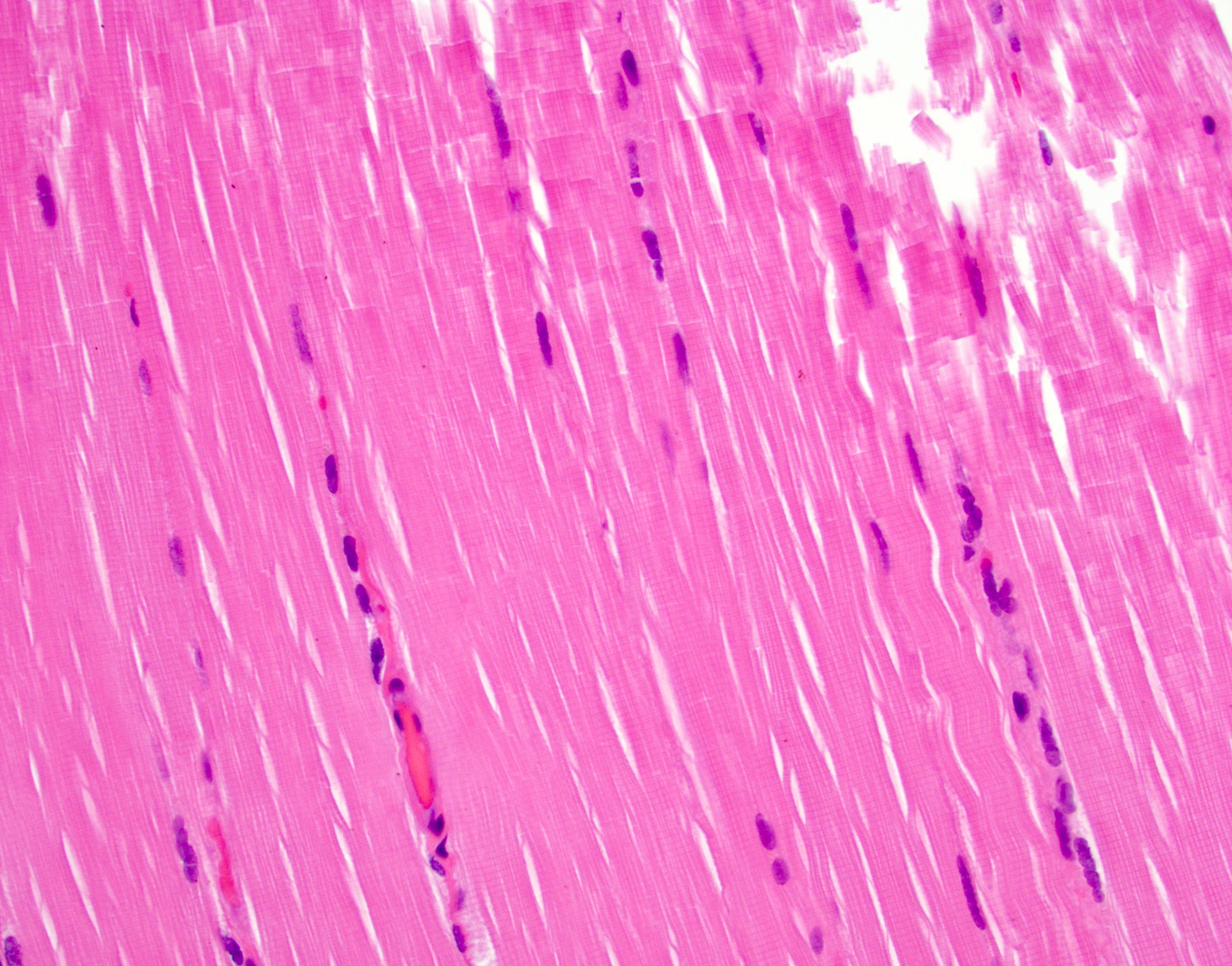
Distinguishing Features on Slide
- Cross-striations (alternating dark A and light I bands) — most distinctive feature
- Multiple peripheral nuclei along fibre edge
- Large fibre diameter compared to smooth/cardiac muscle
- In cross-section: polygonal profiles with peripheral nuclei
- Perimysium and endomysium connective tissue visible between fibres
References
3 sources- 1
Ross MH, Pawlina W. Histology: A Text and Atlas (8th ed.). Wolters Kluwer; 2020.
- 2
Young B, O'Dowd G, Woodford P. Wheater's Functional Histology (6th ed.). Churchill Livingstone/Elsevier; 2014.
- 3
Junqueira LC, Carneiro J. Basic Histology: Text & Atlas (13th ed.). McGraw-Hill; 2013.
Disclaimer: These notes are for educational purposes only and compiled from standard histology textbooks. Clinical interpretation of slides requires a qualified histologist or pathologist.
Ready to test yourself?
Apply what you've learned in the Histology Spotting Test