Large Intestine
Lesson 5 of 16 · Detailed theory + identification points
Points of Identification
4 pointsDetailed Theory
Object: Examination of Histological Slide of Large Intestine
General Overview
The large intestine extends ~1.5 m from the ileocaecal valve to the anus. Its primary functions are water and electrolyte absorption, compaction of faeces, mucus secretion for lubrication, and harboring the gut microbiota. It is divided into caecum, ascending, transverse, descending, and sigmoid colon, rectum, and anal canal.
Key Histological Features
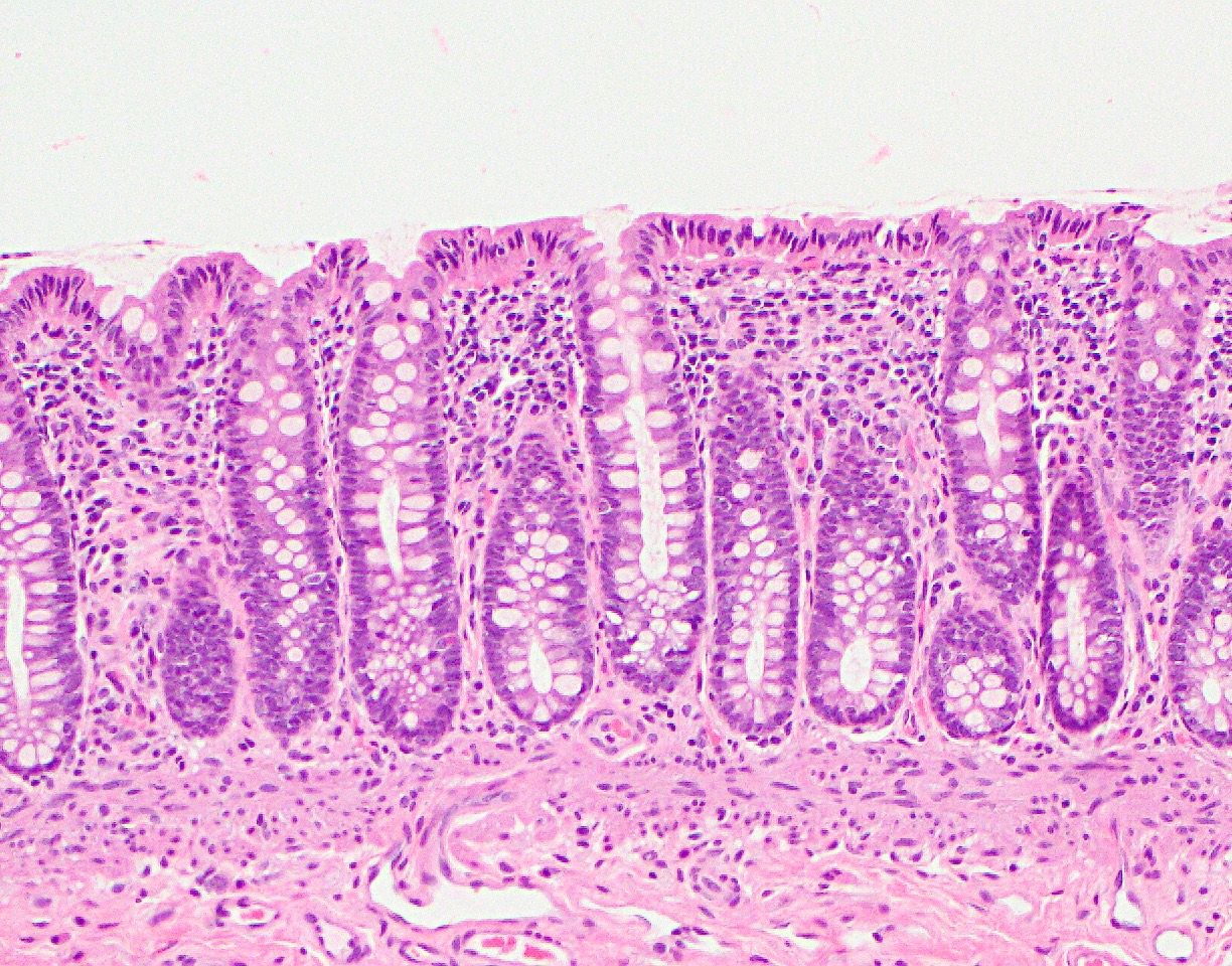
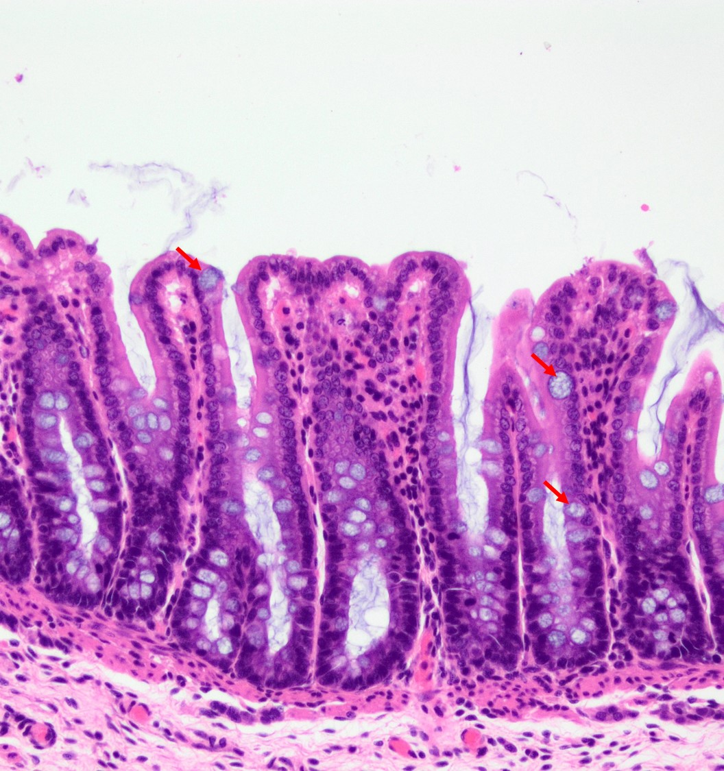
1. Mucosa — No Villi
The large intestine lacks villi — its lining is flat with straight, parallel, tightly packed crypts (intestinal glands). This is the most important distinguishing feature from the small intestine.
- Surface Epithelium: Simple columnar with numerous goblet cells. Goblet cells dramatically increase in number compared to the small intestine — their mucus lubricates the passage of solid faeces.
- Crypts of Lieberkühn: Longer and straighter than in the small intestine, packed with goblet cells. Absorptive cells are fewer; colonocytes absorb water and Na⁺.
- Enteroendocrine Cells: Scattered throughout crypts; secrete peptide YY (PYY) and other hormones regulating motility.
- Lamina Propria: Contains lymphocytes, plasma cells, and abundant diffuse lymphoid tissue forming a protective immune network.
2. Submucosa
Dense irregular connective tissue with blood/lymph vessels and Meissner's plexus. No glands. May contain adipose tissue in the appendices epiploicae.
3. Muscularis Externa — Taeniae Coli
The most distinguishing gross and histological feature of the colon. The outer longitudinal smooth muscle layer is condensed into three bands called taeniae coli, leaving the intervening areas thin. Contraction of taeniae coli shortens the colon and creates sacculations — haustra — visible as outpouchings. Between haustra, semicircular folds are present.
4. Serosa
Contains appendices epiploicae — small fatty projections unique to the colon, visible as fat-filled tags on the serosal surface.
Regional Differences
- Rectum: Longer, less folded crypts; no taeniae coli (complete longitudinal muscle); no haustra.
- Anal Canal: Stratified squamous epithelium in lower portion; internal anal sphincter (smooth muscle); external anal sphincter (skeletal muscle).
Distinguishing Features on Slide
- No villi — flat surface
- Abundant goblet cells — most numerous of any GI segment
- Straight, parallel, long crypts
- Taeniae coli visible as thickened muscular bands
- Abundant lymphoid tissue in lamina propria
- Appendices epiploicae (fat tags) on serosa
References
3 sources- 1
Ross MH, Pawlina W. Histology: A Text and Atlas (8th ed.). Wolters Kluwer; 2020.
- 2
Young B, O'Dowd G, Woodford P. Wheater's Functional Histology (6th ed.). Churchill Livingstone/Elsevier; 2014.
- 3
Junqueira LC, Carneiro J. Basic Histology: Text & Atlas (13th ed.). McGraw-Hill; 2013.
Disclaimer: These notes are for educational purposes only and compiled from standard histology textbooks. Clinical interpretation of slides requires a qualified histologist or pathologist.
Ready to test yourself?
Apply what you've learned in the Histology Spotting Test