

Unit 5: Metabolic Fate of Biomolecules: Carbohydrates, Lipids, Proteins & Amino Acids
Digestion, absorption, and metabolic pathways of carbohydrates, lipids, and proteins — including glycolysis, TCA cycle, β‑oxidation, urea cycle, and heme synthesis.
Prepared by the PharmaWallah student team. Verify results with your textbook or instructor. Report an error · Editorial policy
Unit 5: Metabolic Fate of Biomolecules: Carbohydrates, Lipids, Proteins & Amino Acids
Course Information
- Course Name & Code: Pharmaceutical Biochemistry-I (PHL-309)
- Topic: Metabolic Fate of Biomolecules: Carbohydrates, Lipids, Proteins & Amino Acids
- Credit Hours: 3
Learning Objectives
By the end of this topic, students will be able to:
- Describe the digestion, absorption, and metabolic pathways of carbohydrates, lipids, and proteins.
- Explain glycolysis, the citric acid cycle, and oxidative phosphorylation in energy production.
- Discuss the beta-oxidation of fatty acids and the biosynthesis of lipids and cholesterol.
- Describe the metabolism of amino acids, including urea cycle and heme synthesis.
- Correlate metabolic disorders with their clinical and pharmaceutical significance.
Core Content
PART A: CARBOHYDRATE METABOLISM
5.1 Digestion and Absorption of Carbohydrates
Dietary Sources: Starch, glycogen, sucrose, lactose, and cellulose (indigestible fiber).
Digestion Process:
| Site | Enzyme | Action | Product |
|---|---|---|---|
| Mouth | Salivary α-Amylase | Hydrolysis of α-1,4 glycosidic bonds | Dextrins and maltose |
| Stomach | No carbohydrate digestion | Acidic environment halts amylase activity | – |
| Duodenum | Pancreatic α-Amylase | Continues breakdown of starch/dextrins | Maltose, maltotriose, α-limit dextrins |
| Small Intestine (Brush Border) | Maltase, Sucrase, Lactase | Hydrolysis of disaccharides | Glucose, fructose, galactose (monosaccharides) |
Absorption:
- Glucose and Galactose: Absorbed via SGLT-1 (sodium-dependent glucose transporter) in the intestinal epithelium.
- Fructose: Absorbed via GLUT-5 (facilitated diffusion).
- All monosaccharides are transported into the bloodstream via GLUT-2 at the basolateral membrane and delivered to the liver via the portal vein.
Pharmaceutical Relevance:
- Lactose Intolerance: Deficiency of lactase → osmotic diarrhea, abdominal cramps.
- Sucrase-Isomaltase Deficiency: Causes sucrose intolerance.
- Drug-Drug Interactions: Some drugs (e.g., acarbose) inhibit α-glucosidase to slow carbohydrate absorption in diabetes mellitus.
5.2 Glycolysis (Embden-Meyerhof Pathway)
Definition: The anaerobic breakdown of glucose (6-carbon) into two molecules of pyruvate (3-carbon) with the net production of ATP and NADH.
Site: Cytoplasm of all cells.
Phases of Glycolysis:
Phase 1: Energy Investment Phase (Steps 1-5)
- Glucose is phosphorylated to glucose-6-phosphate (ATP consumed).
- Rearranged to fructose-6-phosphate.
- Phosphorylated to fructose-1,6-bisphosphate (second ATP consumed).
- Cleaved into glyceraldehyde-3-phosphate (G3P) and dihydroxyacetone phosphate (DHAP).
- DHAP is converted to G3P.
Phase 2: Energy Generation Phase (Steps 6-10)
- G3P oxidized to 1,3-bisphosphoglycerate (NADH produced).
- Phosphoryl transfer to ADP → ATP produced (substrate-level phosphorylation).
- 3-phosphoglycerate → 2-phosphoglycerate → Phosphoenolpyruvate (PEP).
- PEP transfers phosphate to ADP → ATP produced (second substrate-level phosphorylation).
- Pyruvate is formed.
Net Equation:
Glucose + 2 NAD⁺ + 2 ADP + 2 Pi → 2 Pyruvate + 2 NADH + 2 H⁺ + 2 ATP + 2 H₂O
Energetics:
- Total ATP consumed: 2
- Total ATP produced: 4 (substrate-level phosphorylation)
- Net ATP gain: 2 ATP (anaerobic); NADH yields additional ATP aerobically via ETC.
Regulation of Glycolysis:
- Key Regulatory Enzymes: Hexokinase/Glucokinase, Phosphofructokinase-1 (PFK-1), Pyruvate Kinase.
- PFK-1 is the most important regulatory enzyme:
- Activators: AMP, Fructose-2,6-bisphosphate.
- Inhibitors: ATP, Citrate, H⁺ (low pH).
Clinical/Pharmaceutical Relevance:
- Diabetes Mellitus: Impaired glycolysis due to insulin deficiency.
- Lactic Acidosis: Excessive anaerobic glycolysis → lactate accumulation.
- Cancer: Warburg effect (increased glycolysis even in presence of oxygen).
- Enzyme Deficiencies: Pyruvate kinase deficiency causes hemolytic anemia.
5.3 Aerobic Breakdown of Glucose
After glycolysis, pyruvate enters the mitochondria for complete oxidation.
A. Pyruvate Dehydrogenase Complex (PDH Complex)
- Converts pyruvate to acetyl-CoA in the mitochondrial matrix.
- Reaction:
Pyruvate + CoA + NAD⁺ → Acetyl-CoA + CO₂ + NADH + H⁺ - Requires: Thiamine (TPP), Lipoic acid, FAD, CoA, NAD⁺.
- Regulation:
- Activators: Insulin, high Ca²⁺ (in muscle).
- Inhibitors: Acetyl-CoA, NADH, ATP (feedback inhibition).
B. Tricarboxylic Acid (TCA) Cycle / Citric Acid Cycle / Krebs Cycle
Definition: A series of enzyme-catalyzed reactions in the mitochondrial matrix that completely oxidizes acetyl-CoA to CO₂ and H₂O, generating NADH, FADH₂, and GTP.
Steps of TCA Cycle:
| Step | Reaction | Enzyme | Products |
|---|---|---|---|
| 1 | Acetyl-CoA + Oxaloacetate → Citrate | Citrate Synthase | Citrate, CoA-SH |
| 2 | Citrate → Isocitrate | Aconitase | Isocitrate |
| 3 | Isocitrate → α-Ketoglutarate | Isocitrate Dehydrogenase | α-KG, NADH, CO₂ |
| 4 | α-Ketoglutarate → Succinyl-CoA | α-KG Dehydrogenase | Succinyl-CoA, NADH, CO₂ |
| 5 | Succinyl-CoA → Succinate | Succinyl-CoA Synthetase | Succinate, GTP (or ATP) |
| 6 | Succinate → Fumarate | Succinate Dehydrogenase | Fumarate, FADH₂ |
| 7 | Fumarate → Malate | Fumarase | Malate |
| 8 | Malate → Oxaloacetate | Malate Dehydrogenase | Oxaloacetate, NADH |
Energetics per Acetyl-CoA:
- 3 NADH → 7.5 ATP (ETC)
- 1 FADH₂ → 1.5 ATP (ETC)
- 1 GTP → 1 ATP
- Total = 10 ATP per Acetyl-CoA
Regulation of TCA Cycle:
- Key enzymes: Citrate Synthase, Isocitrate Dehydrogenase, α-Ketoglutarate Dehydrogenase.
- Activators: Ca²⁺, ADP.
- Inhibitors: ATP, NADH, Succinyl-CoA.
Anaplerotic Reactions: Replenish TCA cycle intermediates (e.g., pyruvate carboxylase converts pyruvate to oxaloacetate).
Pharmaceutical Relevance:
- Fluoroacetate Poisoning: Blocks TCA cycle (inhibits aconitase).
- Arsenic Poisoning: Inhibits α-ketoglutarate dehydrogenase and pyruvate dehydrogenase.
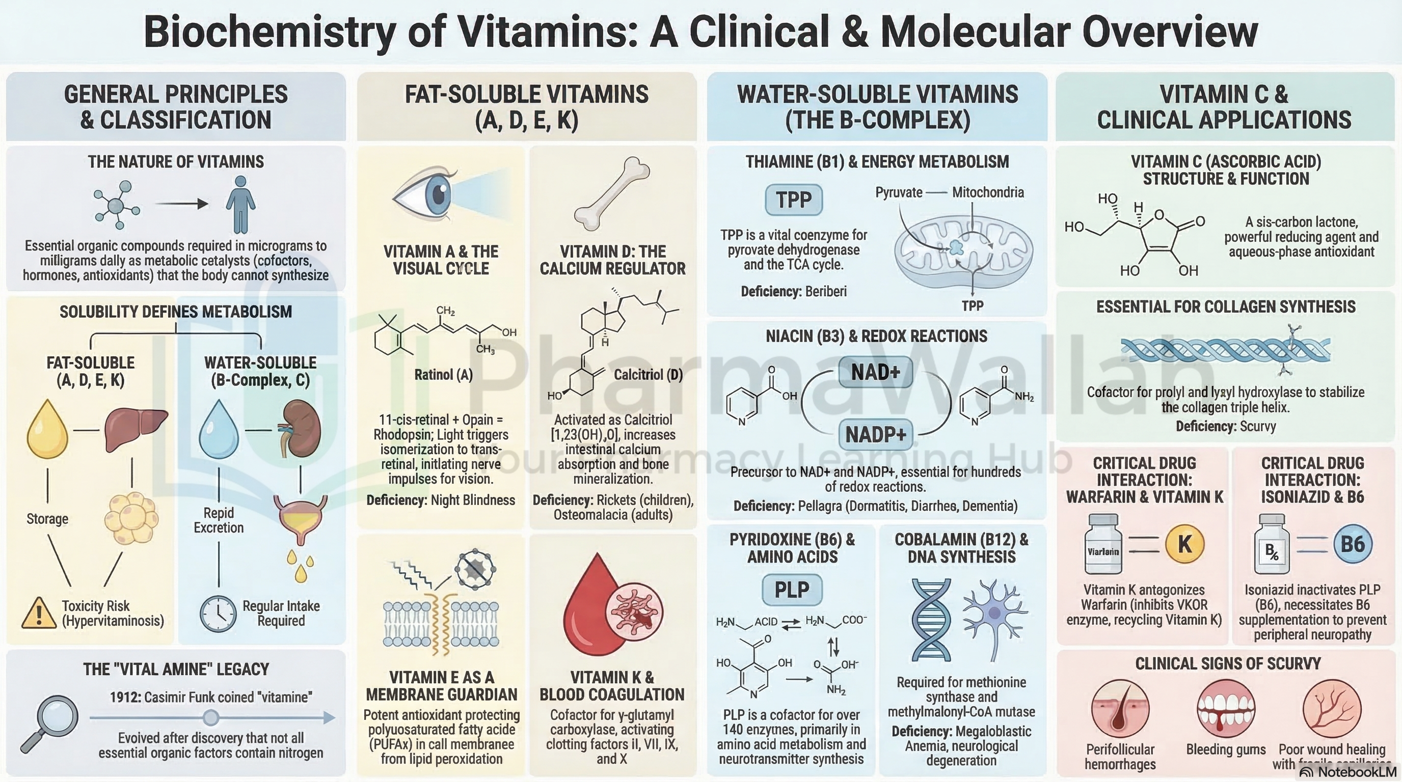
- Vitamin Deficiencies: Beriberi (thiamine deficiency) impairs PDH and TCA.
5.4 Pentose Phosphate Pathway (PPP) / Hexose Monophosphate Shunt
Definition: Alternative pathway for glucose oxidation producing NADPH and pentoses (ribose-5-phosphate) for nucleotide synthesis.
Site: Cytoplasm (liver, adipose tissue, erythrocytes, adrenal cortex, lactating mammary glands).
Phases:
- Oxidative Phase (Irreversible):
- Glucose-6-P → 6-Phosphogluconolactone (G6PD; NADPH produced).
- 6-Phosphogluconolactone → 6-Phosphogluconate.
- 6-Phosphogluconate → Ribulose-5-P + CO₂ (6PGD; NADPH produced).
- Non-Oxidative Phase (Reversible):
- Ribulose-5-P ↔ Ribose-5-P (for nucleotide synthesis).
- Interconversion of sugars to generate glycolytic intermediates (Fructose-6-P and Glyceraldehyde-3-P).
Products:
- NADPH: Reductive biosynthesis (fatty acids, steroids) and antioxidant defense (maintains reduced glutathione).
- Ribose-5-Phosphate: Precursor for nucleotide and nucleic acid synthesis.
Regulation:
- G6PD is the rate-limiting enzyme.
- Activated by: Insulin, NADP⁺.
- Inhibited by: NADPH (feedback inhibition).
Clinical/Pharmaceutical Relevance:
- G6PD Deficiency (Favism): Hemolytic anemia upon oxidative stress (e.g., sulfa drugs, primaquine, fava beans).
- Importance in erythrocytes: only source of NADPH to maintain reduced glutathione.
- Liver: provides NADPH for lipogenesis and cholesterol synthesis.
5.5 Glycogen Metabolism
A. Glycogenesis (Glycogen Synthesis)
- Definition: Synthesis of glycogen from glucose for storage.
- Site: Liver and skeletal muscle.
- Process:
- Glucose → Glucose-6-P (Hexokinase/Glucokinase)
- Glucose-6-P → Glucose-1-P (Phosphoglucomutase)
- Glucose-1-P + UTP → UDP-Glucose + PPi (UDP-Glucose Pyrophosphorylase)
- UDP-Glucose + Glycogen (n) → Glycogen (n+1) + UDP (Glycogen Synthase – rate-limiting enzyme)
- Branching enzyme (α-1,6-glucosidase) creates branch points.
B. Glycogenolysis (Glycogen Breakdown)
- Definition: Breakdown of glycogen to release glucose.
- Site: Liver (releases glucose to blood); muscle (releases glucose-6-P for local energy).
- Process:
- Glycogen (n) + Pi → Glycogen (n-1) + Glucose-1-P (Glycogen Phosphorylase)
- Glucose-1-P → Glucose-6-P (Phosphoglucomutase)
- In liver: Glucose-6-P → Glucose + Pi (Glucose-6-Phosphatase – absent in muscle).
- In muscle: Glucose-6-P enters glycolysis.
Regulation:
- Glycogen Synthase: Activated by insulin; inhibited by epinephrine/glucagon.
- Glycogen Phosphorylase: Activated by epinephrine/glucagon (via cAMP); inhibited by insulin.
Clinical/Pharmaceutical Relevance:
- Glycogen Storage Diseases (GSD):
- Von Gierke's Disease (GSD I): Glucose-6-phosphatase deficiency → severe hypoglycemia, hepatomegaly.
- McArdle's Disease (GSD V): Muscle glycogen phosphorylase deficiency → muscle cramps on exertion.
- Drugs: Glucagon used to treat severe hypoglycemia.
5.6 Gluconeogenesis
Definition: Synthesis of glucose from non-carbohydrate precursors (lactate, amino acids, glycerol, propionate).
Site: Liver (major) and kidneys (minor).
Precursors:
- Lactate (from muscle via Cori cycle).
- Glucogenic amino acids (alanine via glucose-alanine cycle).
- Glycerol (from lipid breakdown).
- Propionate (from odd-chain fatty acids).
Key Reactions (Bypasses of Irreversible Glycolytic Steps):
| Bypass | Glycolytic Enzyme (Irreversible) | Gluconeogenic Enzyme | Reaction |
|---|---|---|---|
| 1 | Pyruvate Kinase | Pyruvate Carboxylase + PEP Carboxykinase (PEPCK) | Pyruvate → Oxaloacetate → PEP |
| 2 | Phosphofructokinase-1 (PFK-1) | Fructose-1,6-Bisphosphatase | Fructose-1,6-BP → Fructose-6-P |
| 3 | Glucokinase/Hexokinase | Glucose-6-Phosphatase | Glucose-6-P → Glucose |
Energetics: Cost: 6 ATP equivalents per glucose molecule synthesized.
Regulation:
- Stimulated by: Cortisol, Glucagon, Epinephrine (fasting state).
- Inhibited by: Insulin (fed state).
Clinical/Pharmaceutical Relevance:
- Diabetes Mellitus: Excessive gluconeogenesis contributes to hyperglycemia.
- Metformin: Decreases gluconeogenesis by activating AMPK.
- Cori Cycle: Lactate from muscle is recycled to glucose in the liver.
- Glucose-Alanine Cycle: Amino groups transported to liver for urea synthesis.
5.7 Energetics of Carbohydrate Metabolism
| Pathway | ATP Yield | Conditions |
|---|---|---|
| Glycolysis (Net) | 2 ATP | Anaerobic |
| Glycolysis + PDH + TCA | ~30-32 ATP | Aerobic (complete oxidation) |
| Pentose Phosphate Pathway | 2 NADPH (≈ 5 ATP equivalents) | Reductive biosynthesis |
| Glycogenolysis (Liver) | – | Releases glucose |
| Glycogenesis | 1 UTP | Requires ATP equivalent |
| Gluconeogenesis | -6 ATP | Net energy consuming |
Complete Oxidation of Glucose:
Glucose + 6 O₂ + 36/38 ADP + 36/38 Pi → 6 CO₂ + 6 H₂O + 36/38 ATP (Old yield = 36/38; modern estimate = 30-32 due to NADH shuttle cost)
PART B: LIPID METABOLISM
5.8 Digestion and Absorption of Lipids
Dietary Sources: Triglycerides (90%), phospholipids, cholesterol esters, fat-soluble vitamins.
Digestion Process:
| Site | Process | Enzymes/Substances | Products |
|---|---|---|---|
| Mouth | Minimal lipid digestion | Lingual lipase | Minor fatty acid release |
| Stomach | Gastric lipase action | Gastric lipase | Some triglycerides → Fatty acids + diglycerides |
| Small Intestine | Emulsification by bile salts | Bile salts (from liver/gallbladder) | Micelles |
| Small Intestine | Enzymatic digestion | Pancreatic lipase, Colipase | Monoglycerides + Free fatty acids (FFA) |
| Small Intestine | Phospholipase A₂ | Lysophospholipids + FFA | |
| Small Intestine | Cholesterol esterase | Cholesterol + FFA |
Absorption:
- Micelles deliver lipids to brush border of enterocytes.
- Lipids diffuse or are transported via transporters into enterocytes.
- Inside enterocytes, lipids are re-esterified to triglycerides, phospholipids, and cholesterol esters.
- Lipids packaged into chylomicrons and secreted into lymph → thoracic duct → bloodstream.
Pharmaceutical Relevance:
- Orlistat: Inhibits pancreatic lipase; used for weight loss.
- Cholestyramine: Bile acid sequestrant; lowers cholesterol.
- Fat Malabsorption: Cystic fibrosis, celiac disease, pancreatic insufficiency.
5.9 Beta-Oxidation of Fatty Acids (β-Oxidation)
Definition: Breakdown of fatty acids into acetyl-CoA units for energy production, occurring in the mitochondrial matrix.
Activation:
- Fatty acids activated to fatty acyl-CoA in cytoplasm using 2 ATP equivalents (ATP → AMP + PPi).
- Carnitine shuttle transports long-chain fatty acyl-CoA into mitochondria.
Steps of β-Oxidation per Cycle:
| Step | Reaction | Enzyme | Products |
|---|---|---|---|
| 1 | Fatty acyl-CoA → Trans-Δ²-enoyl-CoA (dehydrogenation) | Acyl-CoA Dehydrogenase | FADH₂ |
| 2 | Trans-Δ²-enoyl-CoA → L-β-Hydroxyacyl-CoA (hydration) | Enoyl-CoA Hydratase | – |
| 3 | L-β-Hydroxyacyl-CoA → β-Ketoacyl-CoA (dehydrogenation) | β-Hydroxyacyl-CoA Dehydrogenase | NADH |
| 4 | β-Ketoacyl-CoA → Acetyl-CoA + Fatty acyl-CoA (shorter by 2 carbons) | Thiolase | Acetyl-CoA |
Each cycle shortens the fatty acid chain by 2 carbons and produces 1 FADH₂, 1 NADH, and 1 Acetyl-CoA.
Energetics for Palmitic Acid (C16:0):
- Number of cycles = 7
- Products: 8 Acetyl-CoA, 7 FADH₂, 7 NADH
- ATP yield: 8 Acetyl-CoA × 10 = 80 ATP
- 7 FADH₂ × 1.5 = 10.5 ATP
- 7 NADH × 2.5 = 17.5 ATP
- Total = 108 ATP
- Activation cost = 2 ATP
- Net = 106 ATP per palmitate molecule
Regulation:
- Rate-limiting enzyme: Carnitine palmitoyltransferase I (CPT-I) in outer mitochondrial membrane.
- Inhibited by: Malonyl-CoA (a key intermediate of fatty acid synthesis).
- Stimulated by: Fasting, exercise, glucagon, epinephrine.
Clinical/Pharmaceutical Relevance:
- Carnitine Deficiency: Impaired β-oxidation → hypoglycemia, cardiomyopathy.
- Medium-Chain Acyl-CoA Dehydrogenase Deficiency (MCADD): Accumulation of medium-chain fatty acids; hypoketotic hypoglycemia.
- Hypoglycin (Jamaican Vomiting Sickness): Toxin inhibits β-oxidation.
5.10 Biosynthesis of Fatty Acids (Lipogenesis)
Definition: Synthesis of fatty acids from acetyl-CoA, occurring primarily in cytosol of liver, adipose tissue, and lactating mammary glands.
Site: Cytoplasm.
Process Overview:
- Transport of Acetyl-CoA from Mitochondria to Cytosol:
- Acetyl-CoA + Oxaloacetate → Citrate (mitochondria)
- Citrate → Acetyl-CoA + Oxaloacetate (cytosol; via ATP-citrate lyase)
- Activation of Acetyl-CoA to Malonyl-CoA:
- Acetyl-CoA + CO₂ + ATP → Malonyl-CoA (Acetyl-CoA Carboxylase – rate-limiting enzyme; requires biotin).
- Activators: Insulin, Citrate.
- Inhibitors: Palmitoyl-CoA, Glucagon, Epinephrine.
- Fatty Acid Synthase (FAS) Complex:
- Multi-enzyme complex catalyzing sequential addition of 2-carbon units from malonyl-CoA.
- Each cycle: Acetyl (2C) + Malonyl (2C) → 4C chain + CO₂.
- Product: Palmitate (C16:0) after 7 cycles.
Energy Cost for Palmitate (C16:0) Synthesis:
7 Acetyl-CoA + 14 NADPH + 14 H⁺ + 7 ATP → Palmitate + 7 CoA + 14 NADP⁺ + 7 ADP + 7 Pi
Regulation:
- Fed State: Insulin stimulates fatty acid synthesis.
- Fasting State: Glucagon/epinephrine inhibit via phosphorylation of acetyl-CoA carboxylase.
Pharmaceutical Relevance:
- Statins: Inhibit HMG-CoA reductase; reduce cholesterol.
- Fatty acid synthesis inhibitors: potential targets for obesity and cancer therapy.
- Metabolic Syndrome: Dysregulated lipogenesis contributes to fatty liver.
5.11 Biosynthesis of Neutral Lipids
A. Triacylglycerol (Triglyceride) Synthesis
- Definition: Esterification of glycerol-3-phosphate with fatty acyl-CoA to form triglycerides.
- Process:
- Glycerol-3-P + 2 Fatty acyl-CoA → Phosphatidic acid (2 acyl groups).
- Phosphatidic acid → Diacylglycerol (DAG) (dephosphorylation).
- DAG + Fatty acyl-CoA → Triacylglycerol (TAG).
- Source of Glycerol-3-P:
- Liver/kidneys: from glycerol (glycerol kinase).
- Adipose tissue: from dihydroxyacetone phosphate (glycolysis).
B. Phospholipid Synthesis
- Phosphatidic acid → Cytidine diphosphate-diacylglycerol (CDP-DAG).
- Reaction with serine, inositol, or choline yields phosphatidylserine, phosphatidylinositol, and lecithin.
Pharmaceutical Relevance:
- Lecithin: used as emulsifier.
- Phospholipids: essential for liposomal drug delivery.
5.12 Biosynthesis of Cholesterol
Definition: Cholesterol is synthesized from acetyl-CoA via the mevalonate pathway.
Site: Cytoplasm of hepatocytes (mainly); also intestine and adrenal cortex.
Process Overview:
- Acetyl-CoA → HMG-CoA (β-Hydroxy-β-methylglutaryl-CoA): 3 Acetyl-CoA → HMG-CoA (HMG-CoA Synthase).
- HMG-CoA → Mevalonate: HMG-CoA + 2 NADPH → Mevalonate (HMG-CoA Reductase – rate-limiting enzyme).
- Mevalonate → Isopentenyl pyrophosphate (IPP) (via phosphorylation and decarboxylation).
- Condensation of IPP units → Squalene (30-carbon isoprenoid).
- Squalene → Lanosterol (cyclization).
- Lanosterol → Cholesterol (multiple steps).
Regulation:
- HMG-CoA Reductase is the key regulatory enzyme:
- Inhibited by: Cholesterol (feedback), Statins (competitive), Glucagon.
- Stimulated by: Insulin, Thyroid hormone.
Pharmaceutical Relevance:
- Statins (Atorvastatin, Rosuvastatin): HMG-CoA reductase inhibitors; lower serum cholesterol.
- Bile Acid Sequestrants: Bind bile acids, increasing cholesterol excretion.
- Ezetimibe: Inhibits intestinal cholesterol absorption.
- Fibrates: Decrease triglyceride synthesis.
Cholesterol Functions:
- Structural component of cell membranes.
- Precursor for steroid hormones (cortisol, aldosterone, sex hormones).
- Precursor for bile acids and Vitamin D.
- Myelin sheath formation.
PART C: PROTEIN AND AMINO ACID METABOLISM
5.13 Digestion and Absorption of Proteins
Dietary Sources: Meat, fish, eggs, dairy, legumes.
Digestion Process:
| Site | Enzyme | Action | Product |
|---|---|---|---|
| Stomach | Pepsin (activated from Pepsinogen by HCl) | Hydrolysis of peptide bonds | Polypeptides, large peptides |
| Small Intestine | Trypsin, Chymotrypsin, Elastase (pancreatic) | Hydrolysis of internal peptide bonds | Smaller peptides |
| Small Intestine | Carboxypeptidase A/B (pancreatic) | Hydrolysis of C-terminal amino acids | Free amino acids |
| Small Intestine | Aminopeptidases, Dipeptidases (brush border) | Hydrolysis of N-terminal and dipeptides | Free amino acids |
Absorption:
- Free amino acids: absorbed via Na⁺-dependent transporters and H⁺-dependent carriers.
- Small peptides (di/tripeptides): absorbed via PEPT-1 transporters; hydrolyzed to amino acids inside enterocytes.
- Transported to liver via portal circulation.
Pharmaceutical Relevance:
- Protein Malabsorption: Celiac disease, pancreatic insufficiency.
- PEPT-1 Transporters: target for drug delivery (e.g., some antibiotics like cephalexin).
5.14 Metabolism of Essential and Non-Essential Amino Acids
A. General Amino Acid Metabolism
- Transamination:
- Transfer of α-amino group from amino acid to α-keto acid.
- Enzyme: Aminotransferases (Transaminases) – require pyridoxal phosphate (PLP, Vitamin B6).
- Examples:
- Alanine + α-KG ↔ Pyruvate + Glutamate (ALT)
- Aspartate + α-KG ↔ Oxaloacetate + Glutamate (AST)
- Oxidative Deamination:
- Removal of amino group as ammonia (NH₃).
- Glutamate Dehydrogenase: Glutamate + H₂O + NAD(P)⁺ → α-KG + NH₃ + NAD(P)H + H⁺.
- Ammonia converted to urea in liver (urea cycle).
B. Urea Cycle (Ornithine Cycle)
Definition: Series of reactions in liver converting ammonia (NH₃) into urea for excretion.
Site: Liver (mitochondria and cytosol).
Steps:
| Step | Reaction | Location | Enzyme |
|---|---|---|---|
| 1 | NH₃ + CO₂ + 2 ATP → Carbamoyl phosphate | Mitochondria | Carbamoyl Phosphate Synthetase I (CPS-I) |
| 2 | Carbamoyl phosphate + Ornithine → Citrulline | Mitochondria | Ornithine Transcarbamoylase |
| 3 | Citrulline + Aspartate → Argininosuccinate | Cytosol | Argininosuccinate Synthetase (requires ATP) |
| 4 | Argininosuccinate → Arginine + Fumarate | Cytosol | Argininosuccinate Lyase |
| 5 | Arginine + H₂O → Urea + Ornithine | Cytosol | Arginase |
Regulation:
- CPS-I activated by N-acetylglutamate.
Clinical/Pharmaceutical Relevance:
- Urea Cycle Disorders: Hyperammonemia (e.g., Ornithine Transcarbamoylase deficiency).
- Hepatic Encephalopathy: Liver dysfunction → ↑ ammonia → neurotoxicity.
- Drugs: Sodium phenylbutyrate, Sodium benzoate – lower ammonia levels.
C. Metabolism of Specific Amino Acids
| Amino Acid | Metabolic Fate | Deficiencies/Disorders |
|---|---|---|
| Glycine | Heme synthesis, purines, glutathione | Primary Hyperoxaluria (oxalate stones) |
| Phenylalanine | → Tyrosine (phenylalanine hydroxylase) | Phenylketonuria (PKU): mental retardation |
| Tyrosine | → DOPA → Dopamine → Norepinephrine → Epinephrine; melanin, thyroid hormones | Albinism (tyrosinase deficiency), Alkaptonuria |
| Tryptophan | → Niacin (Vitamin B3), Serotonin, Melatonin | Hartnup's Disease |
| Methionine | → S-Adenosylmethionine (SAM); → Homocysteine | Homocystinuria |
| Cysteine | → Glutathione, Taurine, sulfate | – |
| Glutamate | → GABA, Proline, α-KG | – |
| Histidine | → Histamine (histidine decarboxylase) | – |
| Arginine | → Nitric oxide, Creatine, Polyamines | – |
D. Essential vs. Non-Essential Amino Acids
- Essential: Valine, Leucine, Isoleucine, Methionine, Phenylalanine, Threonine, Tryptophan, Lysine, Histidine (in children).
- Non-Essential: Alanine, Aspartate, Glutamate, Serine, etc. – synthesized from carbohydrate metabolism intermediates via transamination.
5.15 Biosynthesis and Catabolism of Haemins and Porphyrins
Definition: Porphyrins are cyclic compounds of four pyrrole rings linked by methene bridges. Heme is protoporphyrin IX + Fe²⁺.
A. Biosynthesis of Heme
- Site: Liver (cytochrome P450), Bone marrow (hemoglobin).
- Steps:
| Step | Reaction | Enzyme | Location |
|---|---|---|---|
| 1 | Glycine + Succinyl-CoA → δ-Aminolevulinic Acid (ALA) | ALA Synthase (rate-limiting; PLP) | Mitochondria |
| 2 | 2 ALA → Porphobilinogen (PBG) | ALA Dehydratase | Cytoplasm |
| 3 | 4 PBG → Hydroxymethylbilane | PBG Deaminase | Cytoplasm |
| 4 | Hydroxymethylbilane → Uroporphyrinogen III | Uroporphyrinogen III Synthase | Cytoplasm |
| 5 | Uroporphyrinogen III → Coproporphyrinogen III | Uroporphyrinogen Decarboxylase | Cytoplasm |
| 6 | Coproporphyrinogen III → Protoporphyrinogen IX | Coproporphyrinogen Oxidase | Mitochondria |
| 7 | Protoporphyrinogen IX → Protoporphyrin IX | Protoporphyrinogen Oxidase | Mitochondria |
| 8 | Protoporphyrin IX + Fe²⁺ → Heme | Ferrochelatase | Mitochondria |
Diagram: Heme Synthesis Simplified
Glycine + Succinyl-CoA | ALA (δ-Aminolevulinic acid) | Porphobilinogen (PBG) | Hydroxymethylbilane | Uroporphyrinogen III | Coproporphyrinogen III | Protoporphyrinogen IX | Protoporphyrin IX | HEME (Protoporphyrin IX + Fe²⁺)
Regulation:
- ALA Synthase is rate-limiting.
- Inhibited by: Heme (feedback).
- Induced by: drugs that induce CYP450 (increase heme demand).
B. Catabolism of Heme
Site: Reticuloendothelial cells (spleen, liver, bone marrow), macrophages.
Process:
- Heme Oxygenase: Heme → Biliverdin + CO + Fe²⁺.
- Biliverdin (green) → bilirubin.
- CO: signaling molecule (vasodilation).
- Fe²⁺: recycled.
- Biliverdin Reductase: Biliverdin → Bilirubin (unconjugated/indirect).
- Transport: Bilirubin bound to albumin → liver.
- Liver Conjugation: Bilirubin + UDP-Glucuronic acid → Bilirubin diglucuronide (conjugated/direct) – glucuronyl transferase.
- Excretion: Conjugated bilirubin → bile → intestine → urobilinogen → stercobilin (feces), urobilin (urine).
Clinical/Pharmaceutical Relevance:
| Condition | Cause | Biochemical Defect | Clinical Features |
|---|---|---|---|
| Jaundice | Elevated bilirubin in blood | Hemolysis, liver disease, bile duct obstruction | Yellow skin/sclera |
| Hemolytic Jaundice | ↑ Unconjugated bilirubin | Excessive RBC breakdown | Pre-hepatic |
| Hepatocellular Jaundice | ↑ Both conjugated/unconjugated | Liver damage (hepatitis, cirrhosis) | Hepatic |
| Obstructive Jaundice | ↑ Conjugated bilirubin | Bile duct obstruction (gallstones) | Post-hepatic |
| Porphyria | Defects in heme synthesis enzymes | Various enzyme deficiencies | Photosensitivity, neuropsychiatric symptoms |
| Acute Intermittent Porphyria | ↓ PBG Deaminase | Accumulation of ALA and PBG | Abdominal pain, neurological symptoms |
Pharmaceutical Importance:
- Porphyrin-based drugs: Photodynamic therapy (PDT) for cancer.
- Bilirubin as diagnostic marker (liver function tests).
- Hematin: used in acute porphyria to suppress heme synthesis.
- Chloramphenicol: causes dose-dependent anemia; may interfere with heme synthesis.
4. Diagrams/Tables
Diagram 1: Overview of Carbohydrate Metabolism
| Hormonal State | Insulin | Glucagon/Epinephrine | Result |
|---|---|---|---|
| Fed State | ↑ | ↓ | Glycolysis ↑, Glycogenesis ↑, Lipogenesis ↑, Glycogenolysis ↓, Gluconeogenesis ↓ |
| Fasting State | ↓ | ↑ | Glycolysis ↓, Glycogenesis ↓, Glycogenolysis ↑, Gluconeogenesis ↑, β-Oxidation ↑ |
Table: Summary of Fatty Acid Oxidation vs. Synthesis
| Feature | β-Oxidation | Fatty Acid Synthesis |
|---|---|---|
| Site | Mitochondria | Cytoplasm |
| Carrier | Acyl-CoA | Acyl carrier protein (ACP) |
| Carbon Units | Acetyl-CoA released (2C) | Malonyl-CoA adds (2C) |
| Redox Cofactors | NAD⁺, FAD (→ NADH, FADH₂) | NADPH (consumed) |
| Direction | Sequential removal | Sequential addition |
| Regulation | Inhibited by Malonyl-CoA | Stimulated by Insulin; inhibited by Glucagon |
Diagram: Heme Synthesis Simplified
Glycine + Succinyl-CoA | ALA (δ-Aminolevulinic acid) | Porphobilinogen (PBG) | Hydroxymethylbilane | Uroporphyrinogen III | Coproporphyrinogen III | Protoporphyrinogen IX | Protoporphyrin IX | HEME (Protoporphyrin IX + Fe²⁺)
5. Exam Angle
Short Questions (2-5 marks)
- Differentiate between glycolysis and gluconeogenesis.
- Write the net equation of glycolysis.
- Name the irreversible enzymes of glycolysis and their bypasses in gluconeogenesis.
- What is the function of the pentose phosphate pathway?
- Define essential fatty acids and give examples.
- Write the sequence of reactions in β-oxidation of fatty acids.
- What is the urea cycle? Name the site and key enzymes.
- Define transamination and oxidative deamination.
- Name the porphyrias and their enzyme defects.
- Why is glucose-6-phosphatase important?
- Differentiate between direct (conjugated) and indirect (unconjugated) bilirubin.
- Explain the Cori cycle.
Long Essays (10+ marks)
- Describe the citric acid cycle in detail with its energetics and regulation. Add a note on its pharmaceutical importance.
- Discuss the digestion and absorption of carbohydrates. Explain glycolysis and its regulation.
- Write a detailed note on β-oxidation of fatty acids, including its energetics and clinical significance.
- Describe the urea cycle and the metabolism of amino acids. Add a note on disorders of the urea cycle.
- Give a detailed account of heme synthesis and its regulation. Discuss the types of jaundice.
- Compare and contrast the metabolism of carbohydrates, lipids, and proteins during fed and fasting states.
Key Points Frequently Tested
- Glycolysis: Irreversible steps (hexokinase, PFK-1, pyruvate kinase); ATP yield; regulation.
- TCA Cycle: Key enzymes; inhibitors; anaplerotic reactions; ATP yield.
- Hormonal Regulation: Insulin vs. Glucagon/Epinephrine on metabolic pathways.
- Clinical Correlations: G6PD deficiency, PKU, MCADD, porphyrias, jaundice, hyperammonemia.
- ATP Yields: Complete oxidation of glucose (~30-32 ATP) and palmitic acid (106 ATP).
- Drug Targets: Statins (HMG-CoA reductase), Orlistat (lipase), Metformin (gluconeogenesis).
6. Summary Box
- Carbohydrate metabolism involves glycolysis (aerobic/anaerobic), TCA cycle, PPP, glycogenolysis, and gluconeogenesis. These pathways are tightly regulated by insulin and glucagon.
- Lipid metabolism includes β-oxidation for energy, fatty acid synthesis (lipogenesis), and cholesterol biosynthesis. β-oxidation of palmitate yields 106 ATP.
- Protein metabolism involves digestion to amino acids, transamination/deamination, and the urea cycle for ammonia detoxification. Essential amino acids must come from the diet.
- Heme synthesis occurs in the liver and bone marrow from glycine and succinyl-CoA. Heme catabolism produces bilirubin, which is conjugated in the liver and excreted via bile.
- Clinical correlations include metabolic disorders (G6PD deficiency, PKU, porphyrias, jaundice), drug targets (statins, orlistat), and diagnostic markers (bilirubin, liver enzymes).
7. References
- Chatterjee, M. N., & Shinde, R. (2011). Textbook of Medical Biochemistry (8th ed.). Jaypee Brothers Medical Publishers.
- Murray, R. K., Bender, D. A., Botham, K. M., Kennelly, P. J., Rodwell, V. W., & Weil, P. A. (2015). Harper's Illustrated Biochemistry (30th ed.). Lange Medical Books / McGraw-Hill Education.
- Nelson, D. L., & Cox, M. M. (2012). Lehninger Principles of Biochemistry (6th ed.). W. H. Freeman.
- Champe, P. C., & Harvey, R. A. (2013). Lippincott's Illustrated Reviews: Biochemistry (6th ed.). Lippincott Williams & Wilkins.
- Peet, A., Lieberman, M. A., & Marks, A. (2012). Marks' Basic Medical Biochemistry (4th ed.). Lippincott Williams & Wilkins.
End of unit 5 of 5
Check what you've learned
5 quick questions from this unit. They're optional: try them if you like, then mark the unit as read and move on.