

Unit 3: Bioenergetics
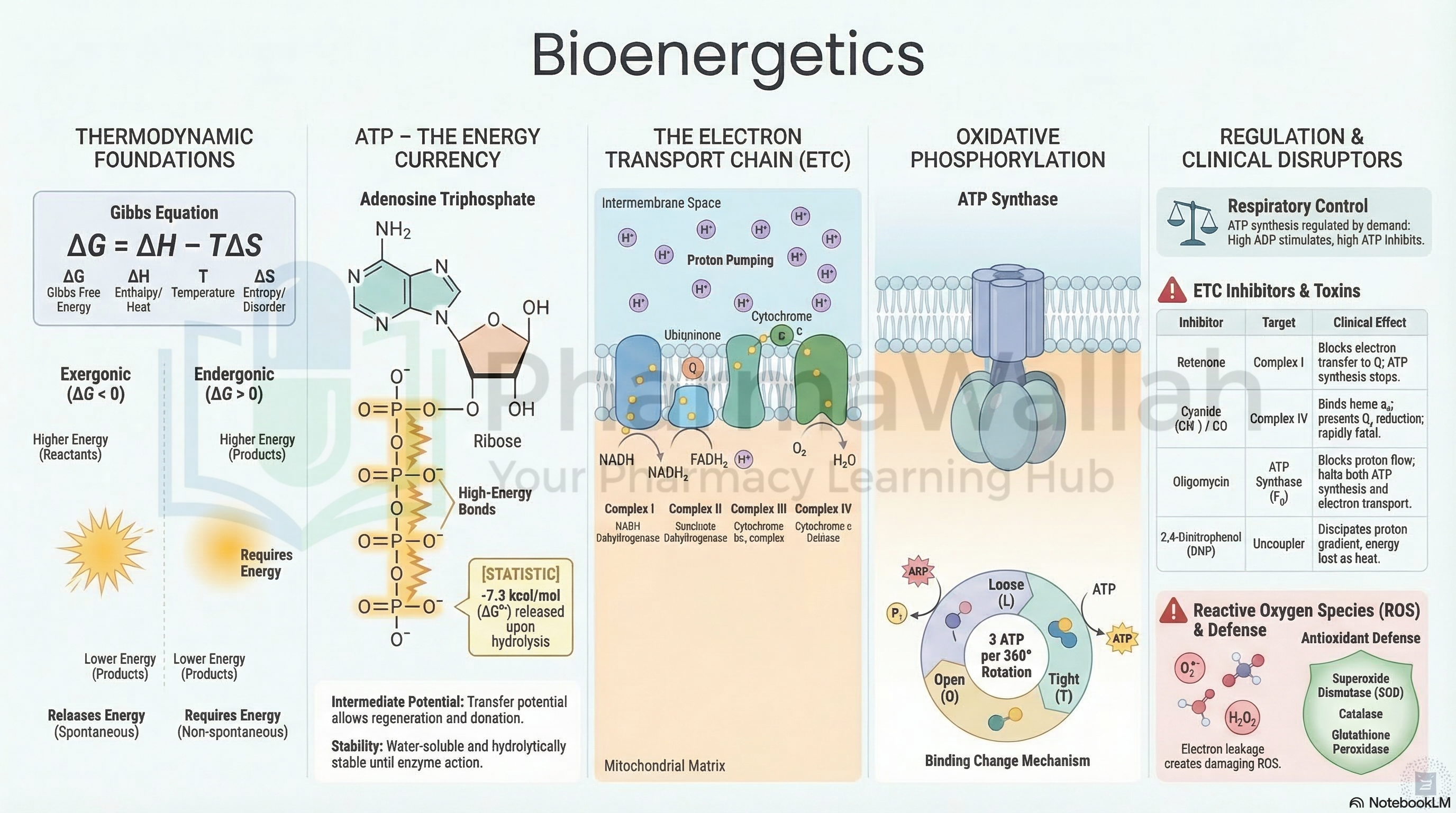
Laws of thermodynamics, free energy, ATP synthesis, oxidative phosphorylation, electron transport chain, and mitochondrial coupling.
Prepared by the PharmaWallah student team. Verify results with your textbook or instructor. Report an error · Editorial policy
Bioenergetics: Principles, Electron Transport Chain, and Oxidative Phosphorylation
Course Information
- Course Name & Code: Pharmaceutical Biochemistry-I (PHL-309)
- Topic: Bioenergetics: Principles, Electron Transport Chain, and Oxidative Phosphorylation
- Credit Hours: 3
Learning Objectives
By the end of this topic, students will be able to:
- Define bioenergetics and explain the fundamental thermodynamic principles governing biochemical reactions.
- Describe the role of ATP as the universal energy currency of the cell.
- Explain the structure and function of the electron transport chain (ETC) complexes in the inner mitochondrial membrane.
- Describe the mechanism of oxidative phosphorylation and chemiosmotic coupling.
- Discuss the pharmaceutical and clinical relevance of ETC inhibitors and uncouplers.
Core Content
3.1 Principles of Bioenergetics
Definition
Bioenergetics is the quantitative study of energy transductions (energy transformations) that occur in living cells. It deals with the concepts of free energy, enthalpy, entropy, and the ways in which cells capture, store, and utilize energy to perform biological work.
Key Thermodynamic Principles
1. First Law of Thermodynamics (Law of Conservation of Energy)
- Energy cannot be created or destroyed; it can only be transformed from one form to another.
- In biological systems: chemical energy (from food) → mechanical work, electrical work, or heat.
2. Second Law of Thermodynamics
- The entropy (degree of disorder) of the universe always increases.
- Living organisms maintain their highly ordered state by increasing entropy in their surroundings (releasing heat).
3. Gibbs Free Energy (G)
- The amount of energy available to do work at constant temperature and pressure.
- Equation:
ΔG = ΔH - TΔS- ΔG = Change in free energy
- ΔH = Change in enthalpy (heat content)
- T = Absolute temperature
- ΔS = Change in entropy (disorder)
| ΔG Value | Type of Reaction | Description |
|---|---|---|
| ΔG < 0 (Negative) | Exergonic | Energy-releasing; spontaneous; favorable |
| ΔG > 0 (Positive) | Endergonic | Energy-requiring; non-spontaneous; unfavorable |
| ΔG = 0 | Equilibrium | No net change; reversible |
ATP: The Energy Currency of the Cell
Definition: Adenosine triphosphate (ATP) is the universal energy carrier in all living cells. It consists of:
- Adenine (nitrogenous base)
- Ribose (pentose sugar)
- Three phosphate groups (α, β, γ)
High-Energy Bonds:
- The phosphoanhydride bonds between phosphate groups are "high-energy" bonds (ΔG°' ≈ -30.5 kJ/mol).
- ATP + H₂O → ADP + Pi (ΔG°' = -30.5 kJ/mol)
- ATP + H₂O → AMP + PPi (ΔG°' = -45.6 kJ/mol)
Why ATP is "High-Energy":
- Electrostatic Repulsion: Negative charges on phosphate groups repel each other.
- Resonance Stabilization: Products (ADP, AMP, Pi) have more resonance structures than ATP.
- Stabilization by Hydration: Products are better solvated by water.
Energy Coupling:
- Exergonic reactions (e.g., catabolism of glucose) drive endergonic reactions (e.g., protein synthesis) via ATP.
- ATP is continuously formed (by phosphorylation of ADP) and broken down (hydrolysis) in a cyclical process.
Types of Biological Work Powered by ATP
| Type of Work | Example |
|---|---|
| Chemical Work | Biosynthesis (macromolecules, lipids, nucleic acids) |
| Mechanical Work | Muscle contraction, cell motility |
| Transport Work | Active transport across membranes (Na⁺/K⁺-ATPase) |
| Electrical Work | Generation of nerve impulses |
| Thermoregulation | Maintenance of body temperature (heat production) |
Other High-Energy Compounds
| Compound | ΔG°' (kJ/mol) | Function |
|---|---|---|
| Phosphoenolpyruvate (PEP) | -61.9 | Glycolysis intermediate |
| 1,3-Bisphosphoglycerate (1,3-BPG) | -49.4 | Glycolysis intermediate |
| Creatine Phosphate | -43.1 | Muscle energy reserve |
| Succinyl-CoA | -33.5 | TCA cycle intermediate |
| Acetyl-CoA | -31.5 | Carrier of acetyl groups |
| ATP | -30.5 | Universal energy currency |
| Glucose-6-Phosphate | -13.8 | Metabolic intermediate |
Redox Potential and Electron Carriers
Definition: Redox potential (E°') is a measure of the tendency of a molecule to accept electrons (reduction potential).
- More negative E°' → Stronger electron donor (reducing agent).
- More positive E°' → Stronger electron acceptor (oxidizing agent).
Important Electron Carriers in Bioenergetics:
| Carrier | Reduced Form | Oxidized Form | Function |
|---|---|---|---|
| NAD⁺ | NADH | NAD⁺ | Carrier of hydride ions (H⁻) in dehydrogenases |
| FAD | FADH₂ | FAD | Carrier of electrons in dehydrogenases |
| Cytochromes | Fe²⁺ | Fe³⁺ | Iron-containing electron carriers in ETC |
| Ubiquinone (CoQ) | CoQH₂ | CoQ | Lipid-soluble electron carrier in ETC |
| FMN | FMNH₂ | FMN | Carrier in Complex I of ETC |
Standard Free Energy Change and Redox Potential Relationship
Equation: ΔG°' = -n F ΔE°'
- n = Number of electrons transferred
- F = Faraday constant (96,485 J/V·mol)
- ΔE°' = Difference in reduction potentials (E°' acceptor - E°' donor)
Significance: The electron flow from more negative to more positive redox potentials is exergonic and can drive ATP synthesis.
3.2 Electron Transport Chain (ETC)
Definition
The Electron Transport Chain (ETC), also known as the respiratory chain, is a series of protein complexes embedded in the inner mitochondrial membrane that transfers electrons from NADH and FADH₂ to molecular oxygen (O₂), creating a proton gradient that drives ATP synthesis.
Location:
- Inner mitochondrial membrane of eukaryotes.
- Plasma membrane of prokaryotes.
Components of the ETC
The ETC consists of four multi-protein complexes (Complex I–IV) and two mobile electron carriers (Coenzyme Q and Cytochrome c).
Complex I: NADH:Ubiquinone Oxidoreductase (NADH Dehydrogenase)
- Function: Transfers electrons from NADH to ubiquinone (CoQ).
- Electron Flow: NADH → FMN → Fe-S clusters → CoQ (→ CoQH₂).
- Protons Pumped: 4 H⁺ per 2 electrons from matrix to intermembrane space.
- Inhibitors: Rotenone, Amytal, Piericidin A, MPTP (causes Parkinsonism).
Complex II: Succinate:Ubiquinone Oxidoreductase (Succinate Dehydrogenase)
- Function: Transfers electrons from FADH₂ (via succinate in TCA cycle) to CoQ.
- Electron Flow: Succinate → FAD → Fe-S clusters → CoQ → CoQH₂.
- Protons Pumped: None.
- Note: This is the only TCA cycle enzyme embedded in the inner mitochondrial membrane.
- Inhibitor: Malonate (competitive inhibitor).
Complex III: Ubiquinone:Cytochrome c Oxidoreductase (Cytochrome bc₁ Complex)
- Function: Transfers electrons from CoQH₂ to cytochrome c.
- Electron Flow: CoQH₂ → Cytochrome b (Fe³⁺) → Fe-S cluster → Cytochrome c₁ → Cytochrome c.
- Protons Pumped: 4 H⁺ per 2 electrons (via Q cycle).
- Inhibitor: Antimycin A (blocks binding of CoQH₂).
Complex IV: Cytochrome c Oxidase
- Function: Transfers electrons from cytochrome c to molecular oxygen (O₂), reducing it to water.
- Electron Flow: 4 Cyt c (Fe²⁺) + O₂ + 8 H⁺ → 4 Cyt c (Fe³⁺) + 2 H₂O + 4 H⁺ (pumped).
- Protons Pumped: 2 H⁺ per 2 electrons (plus 4 H⁺ consumed from matrix for water formation).
- Contains: Cu centers (CuA and CuB) and heme a/heme a₃.
- Inhibitors: Cyanide (CN⁻), Carbon monoxide (CO), Hydrogen sulfide (H₂S), Azide (N₃⁻).
Mobile Electron Carriers:
- Coenzyme Q (Ubiquinone, CoQ): Lipid-soluble, diffuses in the membrane, carries 2 electrons and 2 protons (→ CoQH₂).
- Cytochrome c: Water-soluble protein, transfers 1 electron (Fe³⁺ ↔ Fe²⁺), located in intermembrane space.
Net Reaction:
NADH + H⁺ + ½ O₂ → NAD⁺ + H₂O (ΔG°' = -220 kJ/mol)
3.2.1 The Q Cycle (In Complex III)
Definition: The Q cycle is a mechanism by which Complex III transfers electrons from ubiquinol (CoQH₂) to cytochrome c while simultaneously pumping protons across the membrane.
Process:
- One CoQH₂ binds to the Q₀ site (outer side).
- It transfers 1 electron to cytochrome c (via Fe-S cluster → cyt c₁).
- The other electron is transferred to CoQ at the Qᵢ site (inner side) to form CoQ⁻ (semiquinone).
- A second CoQH₂ binds to Q₀, donating its 2 electrons.
- One electron goes to another cytochrome c; the second electron reduces CoQ⁻ to CoQH₂.
Net Result: 2 CoQH₂ (4 electrons) → 2 CoQ (oxidized), 2 cytochrome c reduced (each with 1 electron), and 4 H⁺ pumped.
Significance: The Q cycle ensures a balanced electron flow and contributes to the proton gradient.
3.3 Oxidative Phosphorylation
Definition
Oxidative phosphorylation is the process by which ATP is synthesized from ADP and inorganic phosphate (Pi) using the energy released by electron flow through the electron transport chain. It occurs in the inner mitochondrial membrane and consists of two coupled processes:
- Electron Transport through the ETC.
- ATP Synthesis by ATP synthase (Complex V).
Chemiosmotic Theory (Peter Mitchell, 1961; Nobel Prize 1978)
Definition: The chemiosmotic theory explains how the electron transport chain drives ATP synthesis. Energy from electron transport is used to pump protons (H⁺) across the inner mitochondrial membrane, creating an electrochemical gradient (proton motive force) that drives ATP synthesis.
Proton Motive Force (Δp):
- Composed of:
- Chemical gradient (ΔpH): Difference in H⁺ concentration across the membrane.
- Electrical gradient (Δψ): Difference in membrane potential (positive outside, negative inside).
- Magnitude: ~180 mV across the inner mitochondrial membrane.
Components of ATP Synthase (Complex V)
| Component | Subunit | Function |
|---|---|---|
| F₀ Domain | a, b, c (c-ring) | Membrane-embedded; forms proton channel; rotates as H⁺ flows through |
| F₁ Domain | α₃β₃γδε (3α, 3β, γ, δ, ε) | Catalytic domain in matrix; synthesizes ATP; β-subunits contain active sites |
| Stator | a, b, δ | Prevents F₁ from rotating with c-ring |
Mechanism of ATP Synthesis (Binding Change Mechanism – Paul Boyer, Nobel Prize 1997)
- Proton Flow: H⁺ flows from intermembrane space to matrix through F₀, causing c-ring to rotate.
- Rotation: The γ subunit (central stalk) rotates within the α₃β₃ hexamer.
- Conformational Changes: The β subunits undergo three conformational states:
- Open (O): Loose binding; releases ATP.
- Tight (T): Binds substrates (ADP + Pi) tightly and catalyzes ATP formation.
- Loose (L): Loose binding; releases ATP.
- ATP Release: As the γ subunit rotates, it forces conformational changes that drive ATP release.
Energy Cost:
- Each ATP synthesized requires 3 H⁺ (4 H⁺ transported into matrix for each ATP in some tissues).
- ATP yield per NADH: ~2.5 ATP (3 H⁺/ATP × 10 H⁺ = 3.33 → ~2.5 ATP in modern textbooks).
- ATP yield per FADH₂: ~1.5 ATP (enters at Complex II → 6 H⁺ → 2 ATP).
Regulation of Oxidative Phosphorylation (Respiratory Control)
- Primarily regulated by ADP availability:
- High ADP → stimulates ETC → more ATP synthesis.
- Low ADP (high ATP) → inhibits ETC (feedback inhibition).
- Uncoupling Proteins (UCPs):
- UCP-1 (brown adipose tissue): Dissipates the proton gradient as heat (thermogenesis).
Stoichiometry of ATP Synthesis
| Pathway | NADH | FADH₂ | ATP Yield |
|---|---|---|---|
| Glycolysis | 2 NADH (cytosolic) | - | ~5 ATP (malate-aspartate shuttle) OR ~3 ATP (glycerol-3-phosphate shuttle) |
| PDH Complex | 2 NADH (per glucose) | - | 5 ATP |
| TCA Cycle | 6 NADH (per glucose) | 2 FADH₂ (per glucose) | 15 + 3 = 18 ATP |
| Substrate-level Phosphorylation | - | - | 2 ATP (glycolysis) + 2 GTP (TCA) = 4 ATP |
| Total (Theoretical Maximum) | - | - | ~32 ATP per glucose (modern estimate) |
Note: Older textbooks give 36–38 ATP; modern estimates account for the cost of transporting NADH into mitochondria, yielding ~30–32 ATP per glucose.
3.4 Inhibitors and Uncouplers of ETC and Oxidative Phosphorylation
A. Inhibitors of the Electron Transport Chain
| Inhibitor | Complex Inhibited | Site of Action | Effect |
|---|---|---|---|
| Rotenone | Complex I | Blocks transfer from Fe-S to CoQ | Prevents NADH oxidation; causes Parkinsonian symptoms (MPTP) |
| Amytal | Complex I | Blocks NADH dehydrogenase | Used experimentally to study ETC |
| Malonate | Complex II | Competitive inhibitor of succinate dehydrogenase | Blocks succinate oxidation |
| Antimycin A | Complex III | Blocks electron transfer from cytochrome b to Fe-S | Complete inhibition of ETC |
| Cyanide (CN⁻) | Complex IV | Binds to heme a₃; blocks O₂ binding | Rapid asphyxiation; antidote = sodium nitrite + thiosulfate |
| Carbon Monoxide (CO) | Complex IV | Binds to heme a₃ (higher affinity than O₂) | Hypoxic tissue damage |
| Hydrogen Sulfide (H₂S) | Complex IV | Blocks cytochrome c oxidase | Cellular hypoxia; antidote = nitrites |
| Sodium Azide (NaN₃) | Complex IV | Binds to cytochrome c oxidase | Blocks ATP synthesis |
B. Uncouplers of Oxidative Phosphorylation
Definition: Uncouplers are compounds that dissociate (uncouple) electron transport from ATP synthesis by dissipating the proton gradient without allowing ATP to be formed. They increase the permeability of the inner mitochondrial membrane to protons.
| Uncoupler | Mechanism | Effect | Clinical/Pharmaceutical Use |
|---|---|---|---|
| 2,4-Dinitrophenol (DNP) | Proton ionophore; transports H⁺ across membrane | Heat production (thermogenesis); formerly used for weight loss (now banned) | Experimental; toxic (hyperthermia, cataracts) |
| Carbonyl Cyanide m-Chlorophenylhydrazone (CCCP) | Protonophore | Used experimentally to study respiration | Research tool |
| Thermogenin (UCP-1) | Endogenous uncoupling protein (brown fat) | Generates heat (non-shivering thermogenesis) | Important in neonatal thermoregulation and hibernation |
| Fatty acids | Activate UCPs | Mild uncoupling | Physiological regulation |
Effect of Uncouplers:
- Increased O₂ consumption (since electron flow continues).
- Decreased ATP synthesis (gradient dissipated).
- Increased heat production.
Significance:
- Malignant Hyperthermia: Uncontrolled muscle thermogenesis due to defective ryanodine receptors; treated with dantrolene.
- Drug-Induced Hyperthermia: Certain drugs (e.g., halothane, succinylcholine) can trigger malignant hyperthermia.
- Mitochondrial Uncoupling: Implicated in obesity, diabetes, and aging.
C. Inhibitors of ATP Synthase (Complex V)
| Inhibitor | Site of Action | Effect | Clinical Use |
|---|---|---|---|
| Oligomycin | F₀ domain (blocks proton channel) | Prevents ATP synthesis; ETC slows (electron flow inhibited) | Experimental; used to study ATP synthase |
| Dicyclohexylcarbodiimide (DCCD) | Binds to c-subunits of F₀ | Blocks proton flow | Research tool |
| Aurovertin | F₁ domain (β-subunit) | Inhibits ATP synthesis | Research tool |
Effect of Oligomycin:
- Prevents proton flow through F₀ → no ATP synthesis.
- The proton gradient builds up and becomes too high → ETC stops (respiratory control).
- This demonstrates the coupling between ETC and ATP synthesis.
3.5 Shuttle Systems for NADH
Problem: NADH generated in the cytoplasm (during glycolysis) cannot directly cross the inner mitochondrial membrane. It must be transported via shuttle systems.
A. Malate-Aspartate Shuttle
- Location: Liver, kidney, cardiac muscle.
- Process:
- Cytosolic NADH reduces oxaloacetate to malate (malate dehydrogenase).
- Malate enters mitochondria via the malate-α-ketoglutarate transporter.
- Mitochondrial malate is oxidized to oxaloacetate, producing NADH.
- Oxaloacetate is transaminated to aspartate; aspartate exits to cytosol.
- Energetics: 1 cytosolic NADH → 1 mitochondrial NADH → ~2.5 ATP.
- This shuttle maintains redox balance and is important in tissues with high oxidative metabolism.
B. Glycerol-3-Phosphate Shuttle
- Location: Skeletal muscle, brain (less active tissues).
- Process:
- Cytosolic NADH reduces dihydroxyacetone phosphate (DHAP) to glycerol-3-phosphate.
- Glycerol-3-P enters mitochondria and is oxidized by mitochondrial glycerol-3-P dehydrogenase (FAD-linked).
- FAD → FADH₂ → CoQ directly (bypasses Complex I).
- Energetics: 1 cytosolic NADH → 1 FADH₂ → ~1.5 ATP.
- This shuttle is less efficient but faster.
Comparison of Shuttles
| Shuttle | Electron Acceptor | ATP Yield | Tissues |
|---|---|---|---|
| Malate-Aspartate | NAD⁺ → NADH | ~2.5 ATP | Liver, heart, kidney |
| Glycerol-3-Phosphate | FAD → FADH₂ | ~1.5 ATP | Skeletal muscle, brain |
Pharmaceutical Significance:
- Metformin: Inhibits mitochondrial Complex I, reducing gluconeogenesis.
- Fructose-1,6-bisphosphate: Used to study shuttle activity.
- Inborn Errors of Metabolism: Defects in shuttle enzymes cause lactic acidosis.
3.6 Reactive Oxygen Species (ROS) and Mitochondrial Damage
Definition: Reactive oxygen species (ROS) are highly reactive molecules derived from oxygen that can damage cellular components.
Sources:
- Complex I and III: Electron leakage during ETC leads to partial reduction of O₂ to superoxide (O₂•⁻).
- Xanthine oxidase, NADPH oxidase in inflammatory cells.
Types of ROS
| Species | Formula | Reactivity |
|---|---|---|
| Superoxide anion | O₂•⁻ | Moderate |
| Hydrogen peroxide | H₂O₂ | Relatively stable |
| Hydroxyl radical | •OH | Highly reactive (most damaging) |
| Peroxynitrite | ONOO⁻ | Highly reactive (nitrosative stress) |
Antioxidant Defense Systems
| Enzyme | Action | Location |
|---|---|---|
| Superoxide Dismutase (SOD) | 2 O₂•⁻ + 2 H⁺ → H₂O₂ + O₂ | Cytoplasm, mitochondria |
| Catalase | 2 H₂O₂ → 2 H₂O + O₂ | Peroxisomes |
| Glutathione Peroxidase (GPx) | 2 GSH + H₂O₂ → GSSG + 2 H₂O | Cytoplasm, mitochondria |
| Thioredoxin Reductase | Reduces oxidized thioredoxin | Various |
Clinical Relevance:
- Mitochondrial Diseases: Mutations in mtDNA → impaired ETC → increased ROS.
- Neurodegenerative Diseases: Parkinson's, Alzheimer's, ALS (oxidative stress).
- Ischemia-Reperfusion Injury: ROS production after restored blood flow.
- Aging: Accumulation of oxidative damage over time.
Pharmaceutical Interventions:
- Antioxidants: Vitamin C, Vitamin E, N-acetylcysteine (NAC).
- ROS Scavengers: Edaravone, MitoQ (mitochondria-targeted antioxidant).
- Prophylaxis: Allopurinol (xanthine oxidase inhibitor) for ischemia-reperfusion injury.
3.7 Mitochondrial Diseases and Pharmaceutical Relevance
Mitochondrial Diseases
| Disease | Defect | Symptoms | Treatment |
|---|---|---|---|
| Mitochondrial Myopathy | Impaired ETC | Muscle weakness, exercise intolerance | CoQ10, Creatine |
| Leigh Syndrome | Complex I, IV, V defects | Neurological degeneration | Supportive care |
| MELAS | Mitochondrial encephalomyopathy, lactic acidosis, stroke-like episodes | Stroke-like episodes, seizures | CoQ10, Carnitine |
| Kearns-Sayre Syndrome | mtDNA deletions | Ophthalmoplegia, heart block | Supportive care |
| LHON | Leber hereditary optic neuropathy | Sudden vision loss | Idebenone |
Pharmaceutical Applications
- Drug Design: Targeting mitochondrial metabolism for cancer therapy (e.g., 2-deoxyglucose, metformin).
- Mitochondrial-targeted antioxidants: MitoQ, MitoE.
- Gene Therapy: Delivering functional mtDNA into mitochondria.
- Mitochondrial Replacement Therapy: "Three-parent IVF" to prevent mtDNA diseases.
4. Diagrams/Tables
Table 1: Summary of ETC Complexes
| Complex | Name | Electron Donor | Electron Acceptor | H⁺ Pumped | Inhibitor |
|---|---|---|---|---|---|
| I | NADH:Ubiquinone Oxidoreductase | NADH | CoQ | 4 | Rotenone, Amytal |
| II | Succinate:Ubiquinone Oxidoreductase | FADH₂ | CoQ | 0 | Malonate |
| III | Ubiquinone:Cytochrome c Oxidoreductase | CoQH₂ | Cyt c | 4 | Antimycin A |
| IV | Cytochrome c Oxidase | Cyt c (Fe²⁺) | O₂ | 2 | Cyanide, CO, H₂S |
| V | ATP Synthase | ADP + Pi | ATP | - | Oligomycin |
Table 2: Comparison of NADH Shuttles
| Shuttle | Location | Electron Acceptor | ATP Yield | Tissues |
|---|---|---|---|---|
| Malate-Aspartate | Liver, heart, kidney | NAD⁺ | ~2.5 ATP/NADH | High oxidative capacity |
| Glycerol-3-Phosphate | Skeletal muscle, brain | FAD | ~1.5 ATP/NADH | Rapid, less efficient |
5. Exam Angle
Short Questions (2-5 marks)
- Define bioenergetics and explain the concept of free energy.
- Why is ATP considered the "energy currency" of the cell?
- List the components of the electron transport chain in order.
- What is the chemiosmotic theory? Explain briefly.
- Differentiate between NADH and FADH₂ in the ETC.
- Name the inhibitors of Complex I, III, and IV.
- What are uncouplers? Give examples.
- What is the Q cycle?
- Explain the malate-aspartate shuttle.
- Calculate the ATP yield from complete oxidation of glucose.
Long Essays (10+ marks)
- Describe the electron transport chain, its components, and the flow of electrons. Add a note on inhibitors of the ETC.
- Explain oxidative phosphorylation and chemiosmotic coupling with reference to ATP synthase.
- Discuss the energetics of glucose metabolism and calculate the total ATP yield.
- Compare and contrast the various shuttle systems for transferring reducing equivalents into mitochondria.
- Write a detailed note on mitochondrial disorders and the role of ETC in disease.
Key Points Frequently Tested
- ATP Yield: ~30–32 ATP per glucose; 106 ATP per palmitate.
- Inhibitors: Rotenone (Complex I), Antimycin A (Complex III), Cyanide/CO (Complex IV), Oligomycin (ATP synthase).
- Uncouplers: DNP, CCCP, thermogenin (UCP-1) — dissipate proton gradient → heat production.
- Redox Potential: Electrons flow from more negative (NADH) to more positive (O₂).
- Proton Motive Force: ~180 mV; drives ATP synthesis via F₀F₁-ATPase.
- Clinical Correlations: Cyanide poisoning (antidote: sodium nitrite + thiosulfate), mitochondrial diseases, ROS and antioxidant defense.
6. Summary Box
- Bioenergetics is the study of energy transformations in cells, governed by thermodynamic principles and driven by redox reactions.
- ATP is the universal energy currency, synthesized from ADP and Pi via exergonic catabolic reactions.
- The electron transport chain (ETC) is a series of four complexes (I–IV) in the inner mitochondrial membrane that transfers electrons from NADH/FADH₂ to O₂, pumping protons to create a gradient.
- Oxidative phosphorylation couples ETC to ATP synthesis via ATP synthase (Complex V), driven by the proton motive force established by the ETC.
- Inhibitors (rotenone, antimycin A, cyanide, oligomycin) and uncouplers (DNP, thermogenin) are key tools for understanding mitochondrial function and have clinical relevance.
- NADH shuttle systems (malate-aspartate and glycerol-3-phosphate) transport cytosolic reducing equivalents into mitochondria with different ATP yields.
- Mitochondrial dysfunction causes diseases (Leigh, MELAS, LHON) and contributes to neurodegeneration, aging, and ischemia-reperfusion injury.
7. References
- Chatterjee, M. N., & Shinde, R. (2011). Textbook of Medical Biochemistry (8th ed.). Jaypee Brothers Medical Publishers.
- Murray, R. K., Bender, D. A., Botham, K. M., Kennelly, P. J., Rodwell, V. W., & Weil, P. A. (2015). Harper's Illustrated Biochemistry (30th ed.). Lange Medical Books / McGraw-Hill Education.
- Nelson, D. L., & Cox, M. M. (2012). Lehninger Principles of Biochemistry (6th ed.). W. H. Freeman.
- Champe, P. C., & Harvey, R. A. (2013). Lippincott's Illustrated Reviews: Biochemistry (6th ed.). Lippincott Williams & Wilkins.
- Peet, A., Lieberman, M. A., & Marks, A. (2012). Marks' Basic Medical Biochemistry (4th ed.). Lippincott Williams & Wilkins.
End of unit 3 of 5
Check what you've learned
5 quick questions from this unit. They're optional: try them if you like, then mark the unit as read and move on.